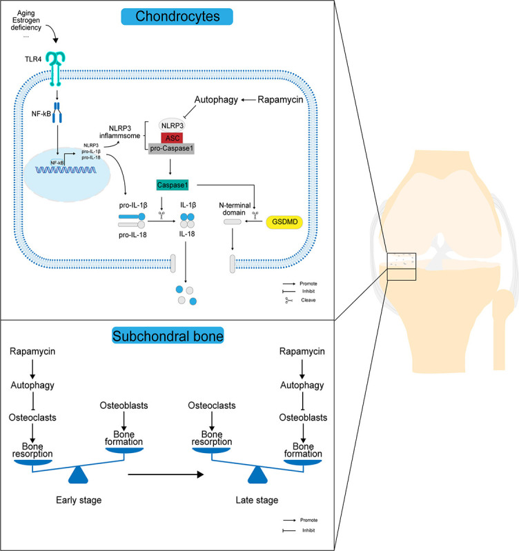
Figure 7.

Scheme of the mechanism by which autophagy affects the progression of OA pathology. In OA chondrocytes, rapamycin activates autophagy while upregulating LC3 expression in chondrocytes, and highly expressed LC3 downregulates NLRP3 inflammasome activation while inhibiting the extracellular release of IL-1β and IL-18 through the NLRP3/caspase-1/GSDMD axis, thus reducing the destruction of articular chondrocyte membranes and the release of inflammatory mediators in the joint cavity. In OA subchondral bone, the activation of autophagy mainly maintains the dynamic balance of subchondral bone resorption and bone formation by inhibiting osteoclast-mediated bone resorption in the early stages and osteoblast-mediated bone formation in the late stages. OA: Osteoarthritis; LC3: Light chain 3; IL: Interleukin; NLRP3: NOD-like receptor protein 3; GSDMD: Gasdermin D; IL-1β: Interleukin-1β; TLR4: Toll-like receptor 4; NF-κB: Nuclear factor kappa B; ASC: Apoptosis-associated specific protein containing a C-termin caspase recruitment domain.
