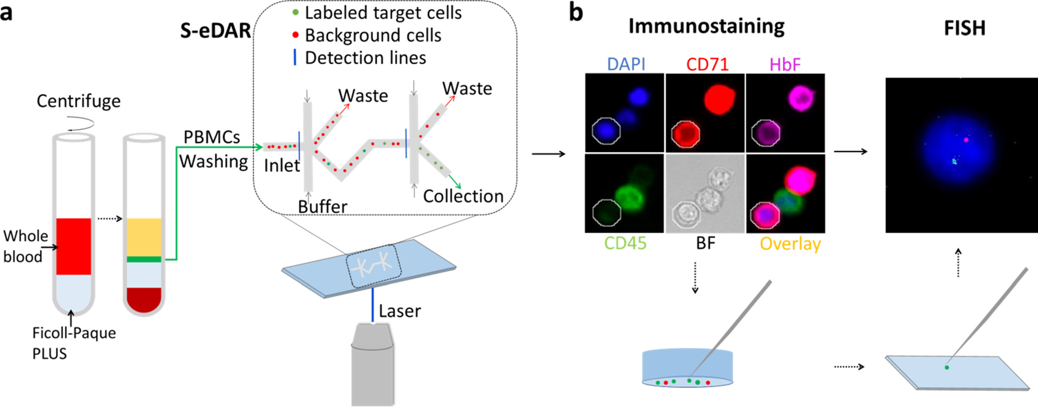
Figure 1.

Workflow for isolation and confirmation of rare cells. (a) Diluted whole blood (red) was layered onto Ficoll-Paque PLUS (gray). After density gradient centrifugation, the thin middle layer (green) containing mononuclear cells between the plasma layer (yellow) and the Ficoll-Paque PLUS layer (light blue) was removed and washed twice and then loaded onto an S-eDAR chip. Labeled target cells were sorted twice at two sorting junctions, accompanied by background cells. (b) The identities of the sorted cells were confirmed by immunostaining and FISH analysis. Once a target cell was confirmed by immunostaining, the cell was picked up by a micromanipulator and transferred onto a slide for FISH analysis.
