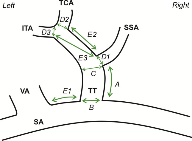
Fig. 1.

Scheme, illustrating collected measurements on an exemplar thyrocervical trunk (TT). SA subclavian artery, VA vertebral artery, SSA suprascapular artery, TCA transverse cervical artery, ITA inferior thyroid artery. A TT length, B TT maximal diameter and ostial area in the start point, C TT maximal diameter and ostial area in the endpoint, D1 maximal diameter and ostial area of the SSA at its origin, D2 maximal diameter and ostial area of the TCA at its origin, D3 maximal diameter and ostial area of the ITA at its origin, E1 shortest distance between the VA and the TT, E2 shortest distance between the SSA and the TCA, E3 shortest distance between the SSA and the ITA
