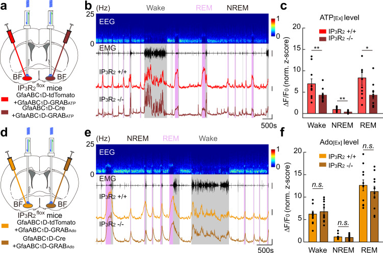
Fig. 6. Suppressing Ca2+ elevation in BF astrocytes decreases extracellular level of ATP but not adenosine.
a Schematic diagram depicting fiber photometry recording of extracellular ATP level in the BF of IP3R2flox mice during the sleep–wake cycle. IP3R2 receptors from astrocytes were knocked out in one hemisphere of the BF via injection of GfaABC1D-Cre, and extracellular ATP level from the two hemispheres of the same mouse was compared. b Top to bottom, EEG power spectrogram, EMG (scale, 2 mV), and GRABATP fluorescence of the two hemispheres, respectively (scale, 1 z-score and 500 s). c Quantification of GRABATP fluorescence in the two hemispheres. n = 10 sessions from 4 mice. Wake, **P = 0.0098, Wilcoxon signed-rank test; NREM, **P = 0.0033, Paired t-test; REM, *P = 0.042, Paired t-test. d–f Same as a–c, respectively, except that extracellular adenosine level was measured. Scale bar in e: EMG, 2 mV; GRABAdo, 1 z-score. In f: n = 11 sessions from 4 mice. Wake, P = 0.48, Paired t-test; NREM, P = 0.27, Wilcoxon signed-rank test; REM, P = 0.34, Paired t-test.

