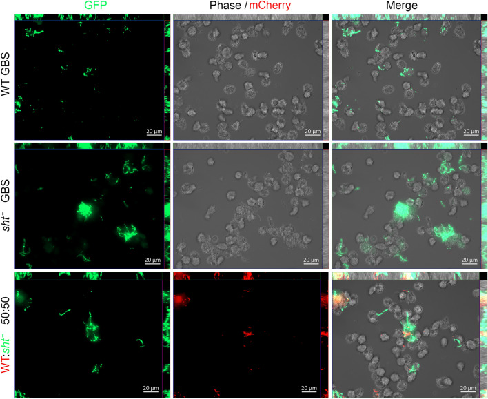
Figure 5.
Visualization of the interaction between human monocytes, WT GBS and Δsht GBS using fluorescence microscopy. The bacteria were tagged with either GFPmut324 or mCherry22 and were used to infect monocytes in single infection assays (one strain of GBS) or mixed infection assays (both strains of GBS tagged with a different fluorophore, used in equal numbers). The images were acquired using a Zeiss AxioImager.M2 microscope and Zen Pro (version 2) software.

