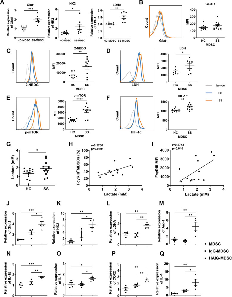
Fig. 4. Peripheral MDSCs of SS patients showed enhanced glycolysis levels, which was promoted by FcγRIIIA activation.
A Peripheral monocytes from HC and SS patients were induced into MDSCs. The mRNA expressions of Glut1, HK2, and LDHA in HC and SS-MDSCs were detected by qPCR (n = 7). B–F Peripheral HLA-DR-CD11b+CD33+ MDSCs were gated to analyze the glucose uptake capacity and protein levels of key factors in glycolytic pathway. Representative flow cytometric analysis and MFI of B Glut1, C 2-NBDG, D LDH, E p-mTOR and F HIF-1α in MDSCs were shown (n = 10 or 7). G The levels of lactate in HC and SS serum were quantified with the Lactate Assay Kit II, H–I and the correlations of lactate levels with FcγRIII+ MDSCs and the FcγRIII MFI of MDSCs in SS patients were analyzed (n = 13). J–Q Peripheral monocytes from HC and SS patients were induced into MDSCs, which were stimulated with IgG and HAIG for 24 h (n = 4). The mRNA expressions of J Glut1, K HK2, L LDHA, M Arg-1, N IL-1β, O IL-6, P COX2, and Q IL-4 were detected by qPCR. All the data are representative of two independent experiments. *p < 0.05, **p < 0.01, ***p < 0.001, ****p < 0.0001.

