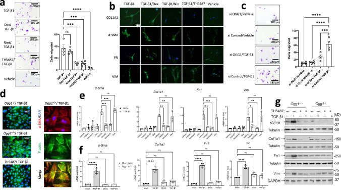
Fig. 1. OGG1-dependent cell migration and fibrotic gene expression.
a Migration of human primary lung fibroblasts (pHLF) post-TGF-β1 induction for 24 h, with significantly more myofibroblast cells appearing than in mock- and inhibitor(s)-treated wells. Data were analyzed using a one-way ANOVA followed by a Dunnett’s post hoc test unless otherwise specified: TGF-β1 vs TGF-β1/TH5487 (P = 0.0001); TGF-β1 vs TGF-β1/Dex (P = 0.6954); TGF-β1 vs TGF-β1/Nin (P = 0.0001); TGF-β1 vs TGF-β1/Veh (P < 0.0001). ns: not significant. Source data are provided as Source data file. Data are representative of 4 independent experiments containing 3 biological replicates (scale bar = 180 μm). Data are presented as the mean ± standard error of the mean (a, c, e, f). b Immunostaining of pHLF cells (green) following 24 h of TGF-β1 treatment, with TH5487 treatment displaying visually reduced levels of collagen (COL1A1), fibronectin (FN), vimentin (VIM), α-smooth muscle actin (α-SMA). Scale bar = 50 μm. Results shown from 3 independent experiments. c Migration of pHLF post-TGF-β1 induction for 24 h, and siRNA transfection targeting OGG1 or scrambled sequence (control). Data were analyzed using a one-way ANOVA followed by a Dunnett’s post hoc test: si Control/TGF-β1 vs si OGG1/TGF-β1 (P = 0.0008); si Control/TGF-β1 vs si OGG1/vehicle and si Control/TGF-β1 vs si Control/vehicle (P < 0.0001). Data are representative of 4 independent experiments containing 3 biological replicates (scale bar = 180 μm). Data are presented as the mean ± standard error of the mean. d Stress fiber formation was seen in response to TGF-β1 in Ogg1+/+, but not Ogg1−/− MF cells (left panel, F-actin). Expression and colocalization of α-SMA (red) with F-actin (green) are shown (right panels, scale bar = 90 μm). Results shown from 3 independent experiments. e The effects of TH5487 on transcription of α-Sma, Fn1, Vim, and Col1A1 in TGF-β1-stimulated MF cells as determined by qRT-PCR. TH5487 (10 μM), significantly decreased mRNA levels of all genes with compiled data representative of 3 independent experiments. Data were analyzed by one-way ANOVA followed by a Dunnett’s post hoc test. f TGF-β1-induced expression of αSma, Col1a1, Fn1, and Vim was decreased in Ogg1−/− but not in Ogg1+/+ MF cells. Data representative of 3 independent experiments. g Immunoblot analysis of TGF-β1-stimulated pHLF cells showing decreased levels of α-SMA, COL1A1, FN1, and VIM levels in OGG1-depleted cells by siRNA. TGF-β1, 2 ng/mL; TH5487, 10 μM; Nintedanib (Nin), 10 μM; Dexamethasone (Dex), 10 μM. Results shown from 3 independent experiments.

