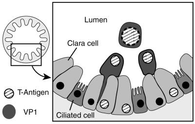
FIG. 4.
In vivo life cycle of polyomavirus in the mouse lung. A schematic of polyomavirus acute replication in the epithelial cells of the mouse bronchiole is shown. Ciliated cells and Clara cells (nonciliated epithelial cells) which make up a majority of the epithelium in mouse bronchioles, are indicated. In the more basal cells, T antigens (early-gene proteins) are expressed, often at very low levels. The cells then transition from a basal location to more apical position near the bronchiolar lumen. During this transition, capsid proteins (late-gene proteins) are expressed and the nuclei of the cells are found apically as well. The cytoplasm of the cells is squeezed from the basement membrane and then dislodged as the cells exfoliate into the bronchiolar lumen (unattached cell). The cells expressing viral proteins (early or late) do not express markers for either ciliated cells or Clara cells, indicating a new differential pathway.

