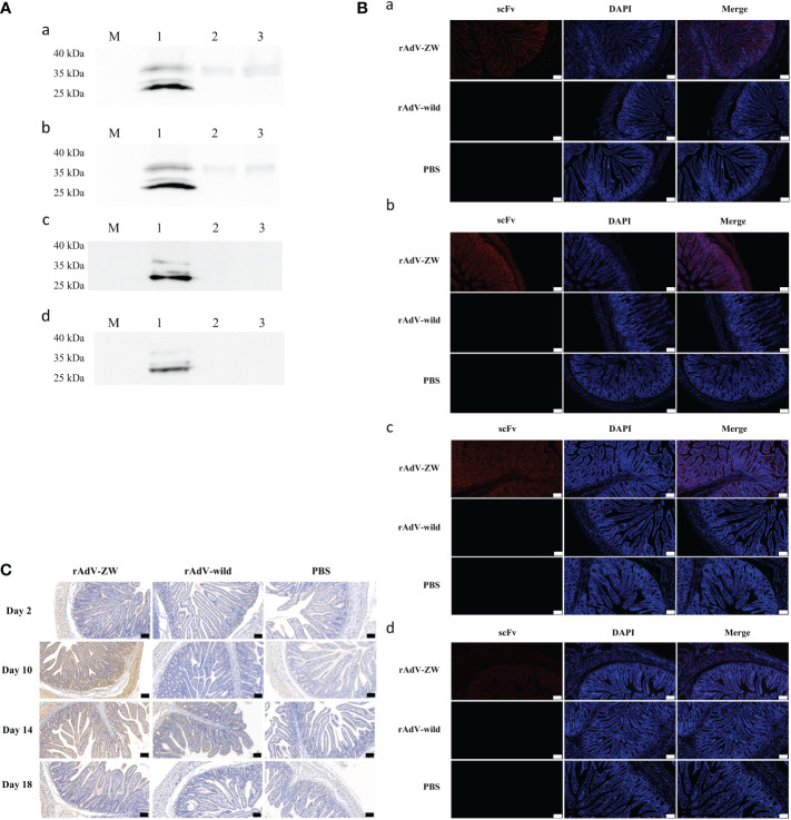
Figure 4.
Jejunal expression of four scFvs. After rAdV-ZW, rAdV-wild-type or PBS oral administration, jejunums were collected on days 2 (Aa, Ba), 10 (Ab, Bb), 14 (Ac, Bc), and 18 (Ad, Bd). (A) Western blotting of scFv expression in jejunum tissue showing several bands at approximately 30-35 kDa. Lanes 1–3, jejunum tissue lysate from piglets administrated rAdV-ZW (lane 1), rAdV-wild-type (lane 2), and PBS (lane 3). (B) Immunofluorescence analysis showing scFv expression in jejunum tissue. Jejunum paraffin sections were incubated with a primary mouse monoclonal anti-HA antibody to detect HA-tagged scFvs, and then stained with DAPI to observe nuclei. Red fluorescence represents scFvs and blue represents nuclei. Scale bar = 100 μm. (C) Immunohistochemical jejunum tissue staining to analyze scFv expression. Brown staining indicates scFvs and blue represents hematoxylin-stained nuclei. Scale bar = 100 μm.

