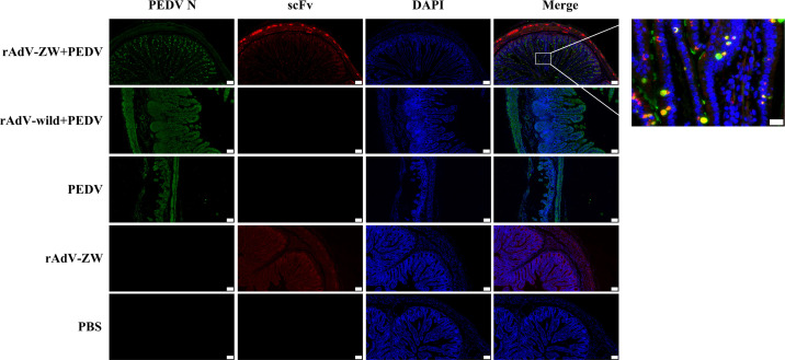
Figure 6.
ScFv and PEDV N protein expression in jejunum by immunofluorescence. Jejunum samples were collected from piglets orally administrated rAdV-ZW after challenge with PEDV. Paraffin sections were stained with FITC, CY3, and DAPI and observed at 100× magnification (left). Scale bar = 100 μm. (Right) (630×) Enlarged framed area in (left). Scale bar = 20 μm. Green represents the PEDV N protein; red represents scFvs, and blue indicates DAPI-stained nuclei. In merged images, orange indicates ZW co-localization (including ZW1-16, ZW3-21, ZW1-41, and ZW4-16) with the PEDV N protein.

