
FIGURE 1.
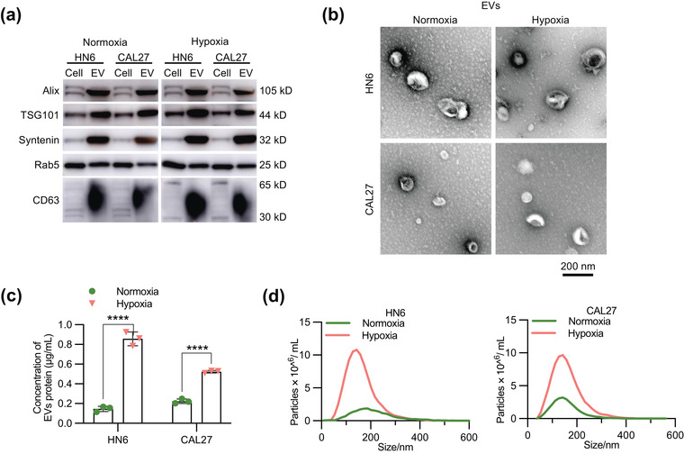
Hypoxia promotes EV secretion. (a) Western blot analysis of EVs purified by ultracentrifugation from cell CM from HN6 and CAL27 cells under normoxia or hypoxia for 48 h. Cells and EVs were blotted for the exosomal markers Alix, TSG101, Syntenin, Rab5 and CD63. (b) Representative electron microscopy images of EVs. Scale bar, 200 nm. (c) The EVs were collected from equal numbers of cells and concentration of EV protein was measured using BCA assay. (d) The size and quantity of normoxic and hypoxic EVs were measured using NTA. EVs were collected from 20 × 106 cells for each group. Statistical analyses were performed using t‐test. ****P‐value < 0.0001. Data are presented as the mean ± S.D.