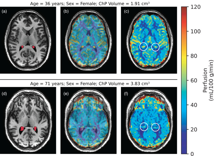
Figure 3.
Choroid plexus (ChP) perfusion-weighted images were produced from pseudo-continuous arterial spin labeling. A 36-year-old, female (a–c) is compared to a 71-year-old, female (d–f). Smaller ChP volume (a: red segment) corresponds with increased perfusion (c: white circle), whereas enlarged ChP volume (d: red segment) corresponds with reduced perfusion (f: white circle). The middle column is included to show adequate co-registration between the perfusion-weighted and T1-weighted images. (ChP: choroid plexus).

