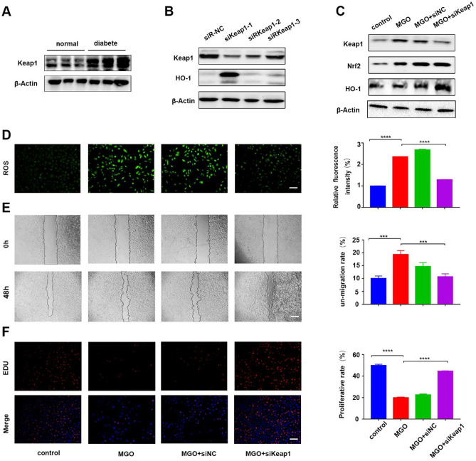
Fig. 1.
Knockdown of Keap1 promotes HUVEC function in an MGO-rich environment. A The expression of Keap1 protein in skin samples from a mouse model of diabetic wounds. B Western blot analysis of the knockdown efficiency of siKeap1. C Western blot analysis of the Keap1/Nrf2/HO-1 pathway in HUVECs in different treatment groups. D Intracellular ROS level of HUVECs in each group. n = 3, ****p < 0. 0001 vs. MGO. Scale bar, 50 μm. E Images of migrated HUVECs in each group. n = 3, **p < 0. 01 vs. MGO. Scale bar, 250 μm. F EDU assay analysis of the proliferation rate of the HUVECs. The proliferative cells and cellular nuclei were stained with red and blue, respectively. n = 3, ****p < 0.0001 vs. MGO by one-way ANOVA. Scale bar, 100 μm. Data is presented as mean ± SD

