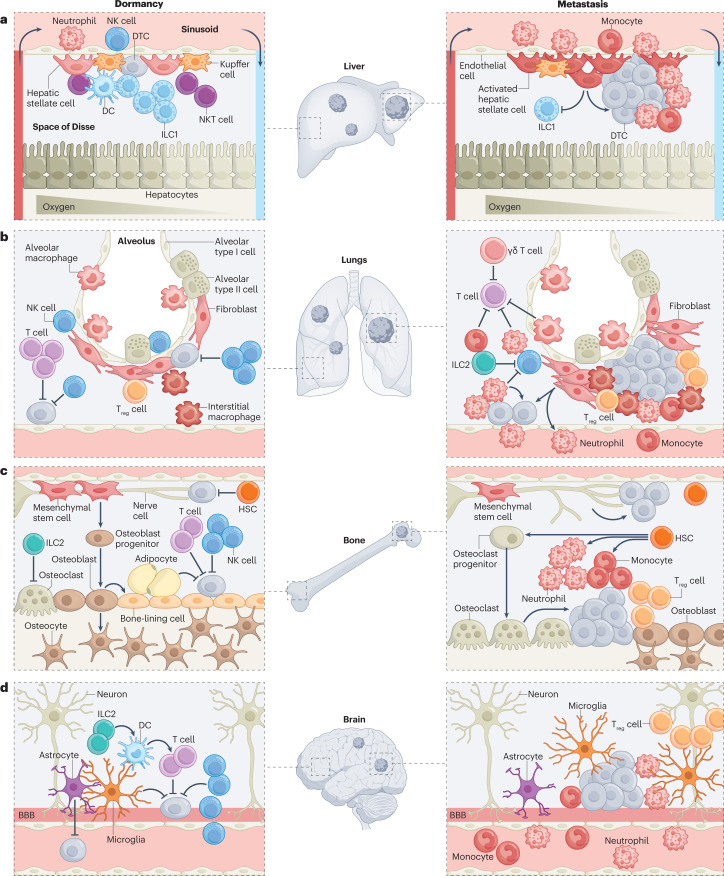
Fig. 2. Site-specific differences of the immune system contribute to differential emergence of metastases within and across common metastatic sites.
The immune system is customized by anatomical site, featuring distinct cell types that are distributed at defined ratios and spatial locations, where they engage in dynamic interactions with diverse non-immune resident cells to constantly safeguard tissue homeostasis. It is the product of these interactions that sets the immune tone for recognition of disseminated tumour cells (DTCs), and makes an ideal locale for DTCs to either be kept dormant or establish metastases within and across different sites. a, In the liver, DTCs migrate from the portal triad vessels to the sinusoid and into the subendothelial space of Disse, where they encounter immune and non-immune resident cells, all positioned in compliance with liver zonation. Liver-resident natural killer (NK) cells (or liver group 1 innate lymphoid cells (ILC1s)) are the main immune gatekeepers of DTC dormancy, whereas activation of hepatic stellate cells precipitates metastasis through suppression of liver-resident NK cell expansion or recruitment of immunosuppressive populations. b, In the lungs, NK cells and T cells are the major immune barriers to metastasis, and their function is countered by diverse immunosuppressive populations, including ILC2s, neutrophils, monocytes, regulatory T cells (Treg cells) and γδ T cells. Tissue-resident macrophages and activated fibroblasts feed the recruitment of these immunosuppressive populations by maintaining a hospitable inflammatory milieu permissive of metastasis. c, The bone is the primary niche for haematopoietic stem cells (HSCs), and is co-opted by DTCs to remain dormant. NK cells and T cells actively survey this niche, in concert with ILC2s that preserve the bone integrity through suppression of bone-resorbing osteoclasts. Conversely, an imbalance towards osteoclast differentiation supports the cycle of bone destruction that triggers dormant DTC reactivation and metastasis initiation. Other awakening factors in the bone include the accumulation of neutrophils, monocytes and Treg cells, as well as enhanced innervation. d, Access to the brain parenchyma is restricted by the blood–brain barrier (BBB), and thus DTC extravasation to the brain takes longer than in other tissues. Once in the brain, DTCs park tightly at the perivasculature, and are actively surveyed and often eliminated by NK cells and T cells, with the latter helped by ILC2-mediated enhanced antigen presentation by dendritic cells (DCs). Microglia, the main immune cell residents of the brain, act in a context-dependent manner, being either suppressive of or permissive of metastatic outgrowth. NKT cell, natural killer T cell.

