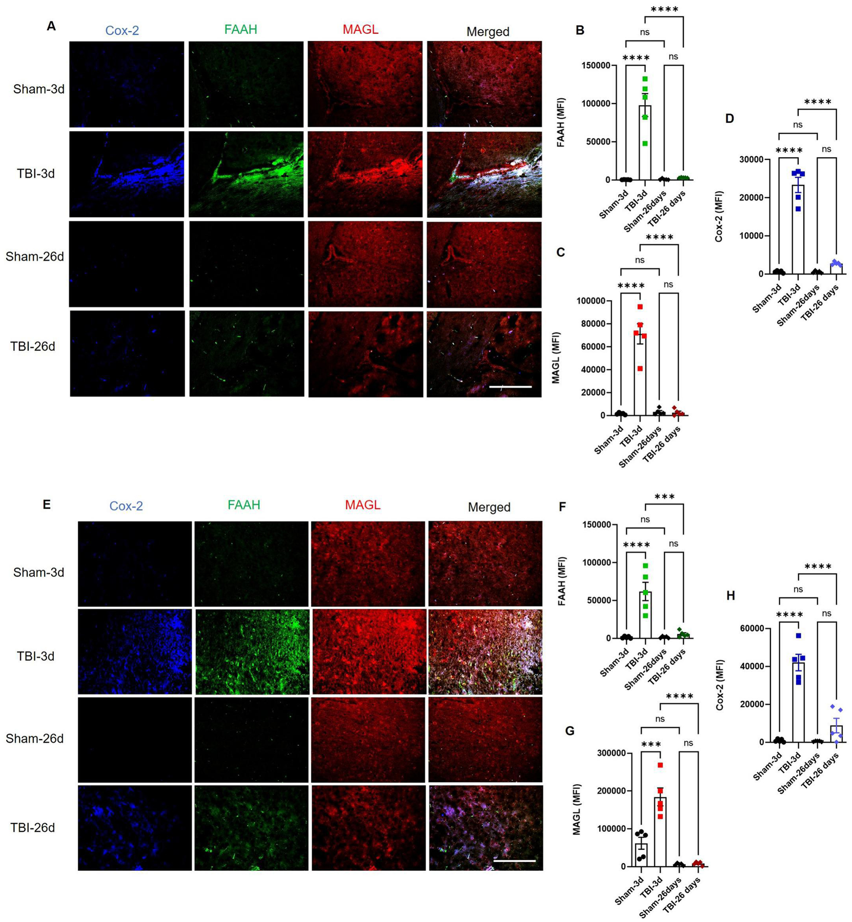
Fig. 3: The CP and pericontusional cortex are sites of acute endocannabinoid metabolism in the TBI mice.

Mouse brain sections were stained for cannabinoid enzymes by immunohistochemistry method. Endocannabinoid metabolizing enzymes FAAH, MAGL and Cox-2 showed higher expressions at 3 days and returned to reduced levels at day 26 post-injury (A-D). As seen at CP, the cortex also shows signs of acute metabolism of 2-AG and AEA as FAAH, MAGL and Cox-2 were highly elevated at day 3 post-TBI with respect to sham (E-H), while only cox-2 remained slightly elevated at day 26th (E, H). Groups were compared by Two-Way ANOVA analysis with Tukey’s post-hoc comparison. Results were calculated as mean fluorescent intensity (MFI) ± SEM. (n=5; ns= not significant; ***p<0.001; ****p<0.0001 vs. sham; Scale bar 125μm).
