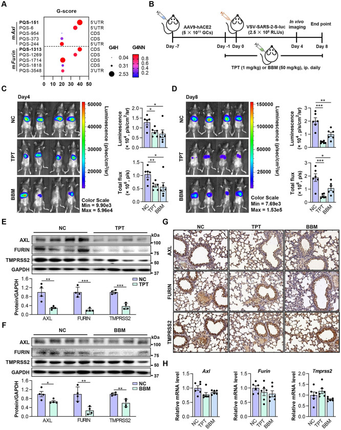
Fig 5. TPT and BBM impede SARS-CoV-2 pseudovirus entry in mice.
(A) RG4 potential of mouse Axl and Furin mRNAs. PQSs conserved between human and mouse are indicated in bold. (B) Schematic of a mouse model for SARS-CoV-2 pseudovirus infection. C57BL/6J mice were transduced intrathoracic with the human ACE2-expressing adenovirus (AAV9-hACE2) and infected with VSV-SARS-2-S-luc after 7 days. Mice received daily peritoneal injections of TPT (1 mg/kg), BBM (50 mg/kg), or vehicle for 9 days from -1 day before VSV-SARS-2-S-luc infection. Following measurements were performed on day 4 and 8 post-infection (n = 6). (C, D) Representative photos of mice on day 4 (C) and 8 (D) post-infection (n = 6). Relative levels of bioluminescence are shown in pseudocolours, with blue and red representing the weakest and strongest photon fluxes, respectively. Photon emission from each mouse was quantified as luminescence (p/s/cm2/sr) and total flux (p/s) post imaging, respectively. (E, F) Protein levels of AXL, FURIN and TMPRSS2 in lungs of VSV-SARS-2-S-luc-infected mice treated with TPT (E) or BBM (F) (upper panel) (n = 4). ImageJ quantification of the target/GAPDH ratio is shown (bottom panel). (G) IHC staining for AXL, FURIN and TMPRSS2 in paraffin lung sections. Scale bars: 50 μm. (H) mRNA levels of Axl, Furin, and Tmprss2 in lungs of VSV-SARS-2-S-luc-infected mice treated with TPT or BBM (n = 6). Data are shown as mean ± SEM, n = 4–6. *p < 0.05, **p < 0.01, ***p < 0.001 (Two-tailed Student’s t test). Fig 5B was created using Microsoft PowerPoint and Adobe Illustrator.

