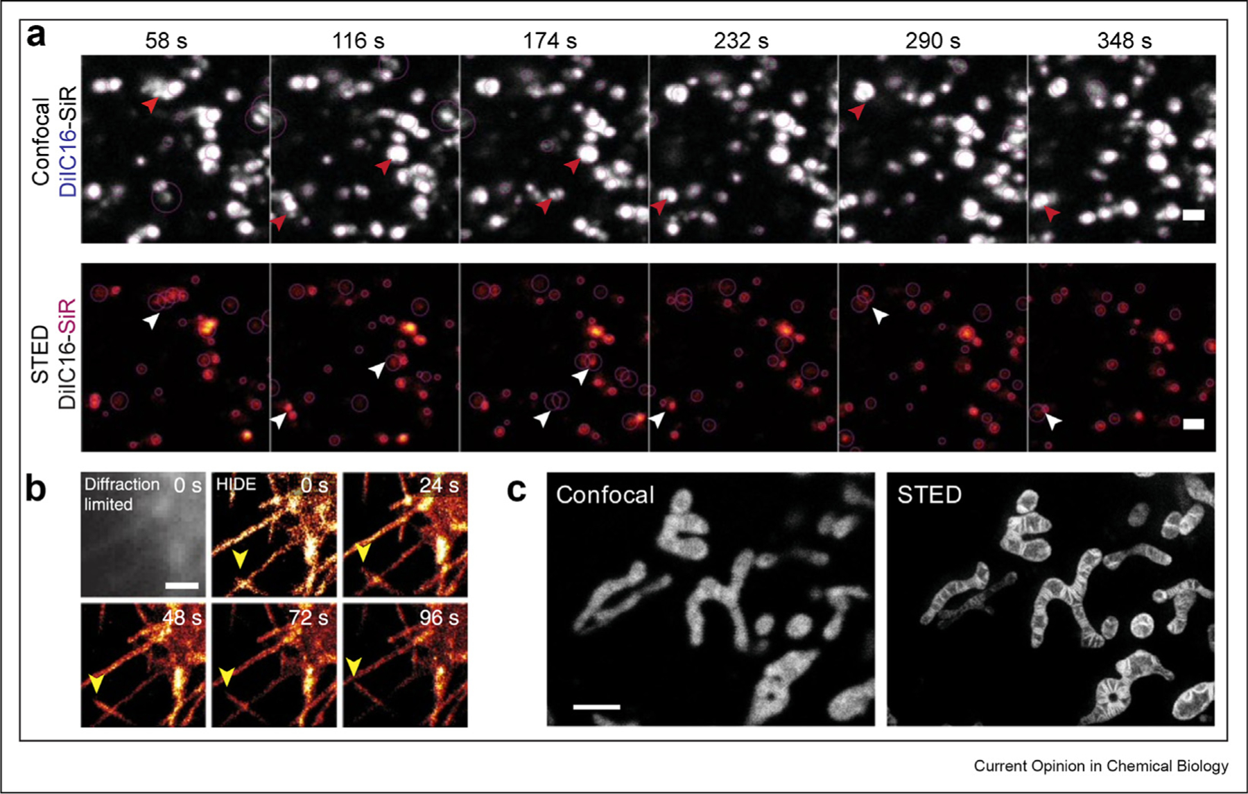
Figure 3. Membrane-targeting small-molecule probes for super-resolution microscopy of organelles.

(a) Visualizing endosomal motility defects with DiI-C16-SiR in wild-type fibroblasts from patients with Niemann-Pick C disease; scale bar: 1 μm. Reprinted by permission from Springer Nature Customer Service Centre GmbH: Nature Chemical Biology, Endosome motility defects revealed at super resolution in live cells using HIDE probes, Gupta et al., 2020 (b) SMLM time-lapse images of plasma membrane filopodia acquired using DiI-HMSiR; scale bar: 1 μm. Reprinted by permission from Springer Nature Customer Service Centre GmbH: Nature Biotechnology, Long time-lapse nanoscopy with spontaneously blinking membrane probes, Takakura et al., 2017 (c) Confocal vs. STED images of the mitochondria in HeLa cells using MitoPB Yellow; scale bar: 2 μm. Reprinted from Wang C et al.,: A photostable fluorescent marker for the super-resolution live imaging of the dynamic structure of the mitochondrial cristae. Proc Natl Acad Sci U S A 2019, 116:15817–15822. SMLM, single-molecule localization microscopy; STED, stimulated emission depletion.
