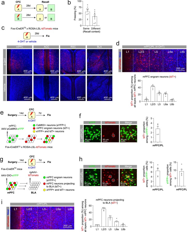
Extended Data Fig. 1. Labeling of mPFC engram neurons and their proportions among all CaMKII+ neurons and among all BLA projectors.
(a) Mice were fear conditioned in Context A or B and tested for remote memory recall in the same (9 mice) or different context (9 mice). (b) Quantification of freezing behavior during remote memory recall as in (a). Mice tested in the same contexts tended to display more freezing behavior than mice tested in different contexts (p = 0.07, unpaired t-test). (c) Neurons active during CFC (engram neurons) were labeled with tdTomato (tdT). Images show tdT+ neurons (red) in mPFC, caudal anterior cingulate cortex (ACC), retrosplenial cortex (RSC), and basolateral amygdala (BLA) in mice that received 4-OHT but not vehicle injection after CFC (Blue: Nissl stain). (d) Top: tdT+ neurons (red) in different mPFC/PL layers in 4-OHT-injected mice in (c). Bottom: proportion of tdT+ neurons in each mPFC/PL layer among all tdT+ neurons (6 mice). (e) Experimental setup for (f). CaMKII+ mPFC pyramidal neurons expressed eYFP, whereas neurons active during CFC expressed tdT. (f) Left: eYFP+ (green) or tdT+ (red) mPFC/PL neurons. A both eYFP+ and tdT+ neuron is circled. Right: proportion of tdT+ neurons among all eYFP+ mPFC neurons (5 mice). (g) Experimental setup for (h)-(i). mPFC neurons projecting to BLA (BLA projectors) were retrogradely labeled with tdT. mPFC neurons active during CFC expressed eYFP. (h) Left: tdT+ (red) or eYFP+ (green) mPFC/PL neurons. Both tdT+ and eYFP+ neurons are circled. Middle: proportion of tdT+ neurons among all eYFP+ mPFC neurons. Right: proportion of eYFP+ neurons among all tdT+ mPFC neurons (5 mice). (i) Left: BLA projectors (tdT+, red) in different mPFC/PL layers. Right: proportion of BLA projectors in each mPFC/PL layer among all BLA projectors in mPFC (5 mice). Data are presented as the mean ± SEM.

