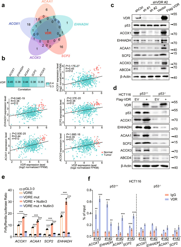
Fig. 2. p53 is associated with VDR to promote the expression of genes involved in peroxisomal β-oxidation.
a Venn diagram showing the numbers of overlapping TFs among the peroxisomal FAO rate-limiting enzyme genes. b Correlation between the expression levels of VDR and ACOX1, EHHADH, ACAA1, SCP2, ACOX3 in 41 normal (orange) and 471 tumor (green) COAD samples, as determined by Pearson’s r analysis. c Protein expression of ACOX1, EHHADH, ACAA1, SCP2, ACOX3, and ABCD4 in HCT116 cells with VDR inhibition. d Western blot analysis of ACOX1, EHHADH, ACAA1, SCP2, ACOX3 and ABCD4 expression in HCT116 p53+/+ or p53−/−cells expressing VDR. e Dual-luciferase assays in HEK293T cells transfected with the indicated plasmids or treated with Nutlin3 (10 μΜ), n = 3. f qPCR ChIP analyses of VDR binding to ACOX1, EHHADH, ACAA1, and SCP2 promoter regions in HCT116 p53+/+ or p53−/−cells, n = 3. Data were presented as mean ± SD. **P < 0.01, ***P < 0.001.

