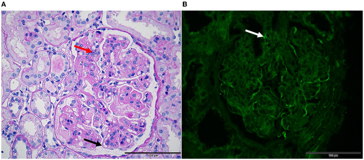
Figure 2.
Renal pathology in mixed cryoglobulinemia. Light microscopy ( × 10). (A) PAS stain shows membranoproliferative glomerulonephritis characterized by nodular lobulated changes in glomeruli, segmentally thickened and double-contoured appearance of the glomerular basement membrane (black arrow), diffuse intracapillary cell accumulation, and inflammatory cell infiltration (red arrow). (B) Immunofluorescence studies showing Immunoglobulin G deposit in vascular loops with line-like (white arrow).

