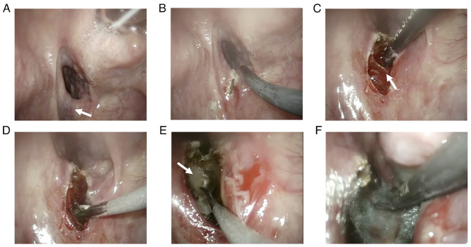
Figure 4.
Intraoperative photographs. (A) The field of view for identifying the cricopharyngeal muscle. The arrow in the figure indicates the cricopharyngeal muscle as a submucosal ridge on the posterior wall of the esophageal inlet. (B) Mucosal incision. (C) Detachment of the cricopharyngeal muscle from the surrounding tissue. The arrow in the figure indicates the cricopharyngeal muscle. (D) Cutting of the cricopharyngeal muscle. (E) Buccopharyngeal fascia visible after cutting the cricopharyngeal muscle. The arrow in the figure indicates the buccopharyngeal fascia. (F) Wound protection with a polyglycolic acid sheet after resection.

