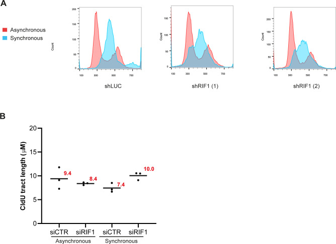
Figure S2. Impact of RIF1 depletion on replication forks dynamic upon synchronization in G1 and release into S-phase.
(A) Flow cytometry analysis of cells depleted or not for RIF1 in asynchronous or synchronous conditions (18-h thymidine block followed by 2-h release). DNA was stained using propidium iodide. (B) This graphic representation is showing the average of the three independent experiments from Fig 2F.

