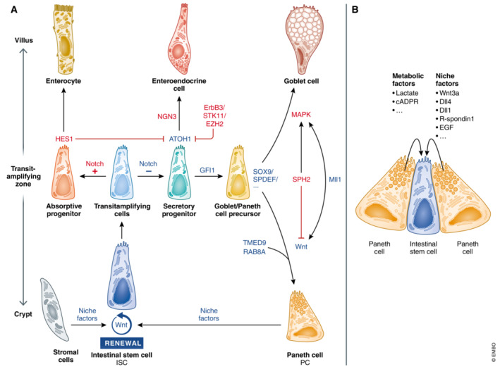
Figure 1. Intestinal cell differentiation pathways and signals.

(A) Wnt and Notch signals control ISC renewal and differentiation. PCs and for example, pericryptal stromal cells support the stemness of ISCs by providing niche factors. ISCs in the lower part of the crypt give rise to a larger pool of transit‐amplifying cells in the transit‐amplifying zone. Differentiation from transit‐amplifying cells towards secretory or absorptive progenitors is initially controlled by Notch signaling. Cells that receive Notch signals express HES1, a negative regulator of ATOH1, leading to differentiation into absorbing enterocytes. Cells devoid of Notch express ATOH1, a hallmark of secretory cells. GFI1 acts downstream of ATOH1 to select for goblet cell and PC differentiation, as it represses neurogenin 3 (NGN3), a transcription factor involved in enteroendocrine cell differentiation. ErbB3, STK11, and EZH2 influence PC differentiation by reducing ATOH1 levels and affect the numbers and/or location of mature PCs. SOX9 and SPDEF are involved in PC and goblet cell differentiation. Reduced MAPK signaling and increased Wnt signaling (influenced by Wnt regulating factors, e.g., RAB8A, TMED9, SPH2, and Mll1) favor differentiation into PCs instead of goblet cells. (B) PCs support ISCs by providing niche and metabolic factors. Blue – factors that promote PC differentiation, red – factors that inhibit PC differentiation.
