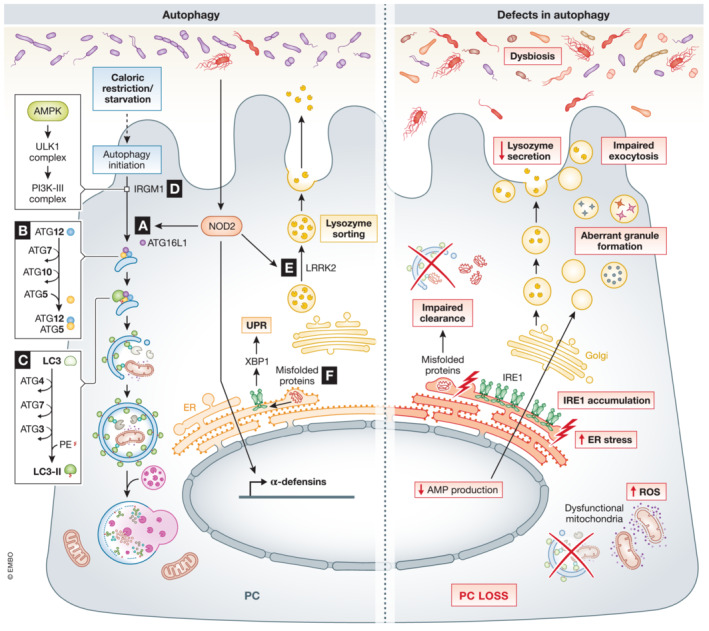
Figure 4. Role of autophagy and ER in PCs.

(A) Bacterial activation of NOD2 initiates direct autophagy by recruiting ATG16l1. Defects in Nod2 and/or Atg16l1 have been related to a decrease in AMP production, impaired exocytosis, accumulation of IRE1, and bacterial translocation. (B) In PCs, ATG5 binds ATG16L1 and ATG12, which is important in the early stages of autophagy, catalyzing the microtubule‐associated protein light chain 3 (LC3) lipidation. Specific deletion of Atg5 in PCs has been associated with accumulation of ROS and increased ER stress due to impaired clearance of dysfunctional mitochondria. C) ATG4 and ATG7 form a complex with ATG3, responsible for the biogenesis of the autophagosome by determining the site of LC3 lipidation. Dysfunction in autophagy, aberrant granule formation, and defects in AMP production and secretion have been observed in Atg4b KO and Atg7 ΔIEC mice. D) IRGM1 plays a direct role in organizing the autophagy process. IRGM1 can initiate the phosphorylation cascade that activates Unc‐51 like autophagy activating kinase 1 (ULK1) and Beclin1 (an autophagy regulator part of the PI3K‐III complex), which promotes autophagy. Defects in Irgm1 have been linked to alterations in PC numbers and location, abnormal PC morphology and aberrant granules. E) LRRK2, together with RIP2 and RAB2A, coordinates lysozyme sorting after recruitment by bacterial‐activated NOD2. Dysfunctions in Nod2 and/or Lrrk2 result in compromised lysozyme secretion. F) Misfolded or unfolded proteins are recognized by IRE1, which by unconventional splicing generates XBP1, activating UPR response. Specific deletion of Xbp1 in PCs leads to PC dysfunction, a condensed ER and abnormal secretory granules. Black arrows – pathways that are active in PCs, red arrows – “up‐ or downregulation” of factors that negatively affect PCs function.
