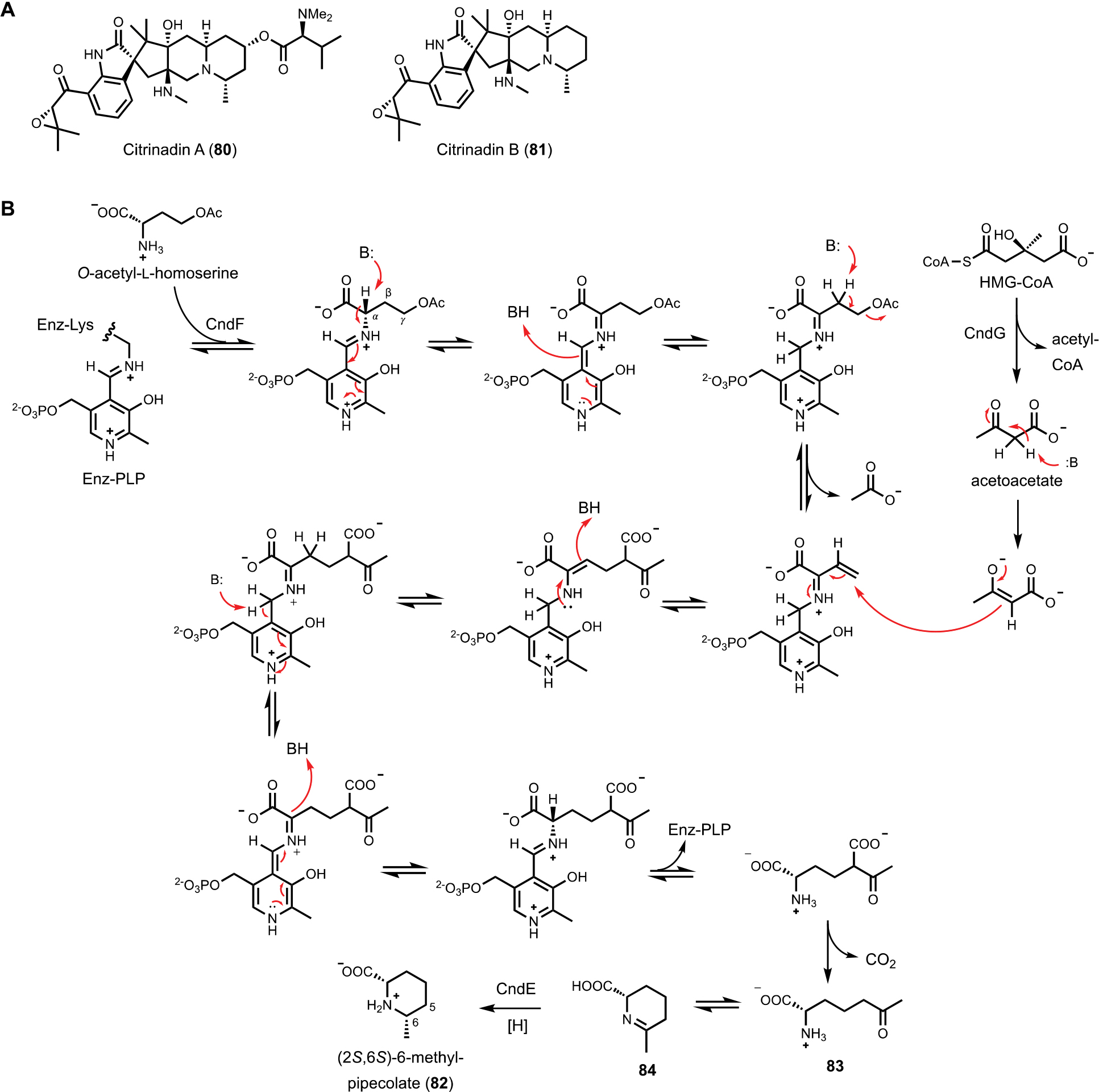
Figure 13.

(A) Structure of citrinadins. (B) Biosynthetic pathway of forming (2S,3S)-6-methylpipecolate. The mechanism of 𝛾-substitution catalyzed by CndF was proposed. O-acetyl-L-homoserine was first bound to PLP as the external aldimine. After eliminating acetate, acetoacetate generated from HMG-CoA by HMG-CoA lyase then attacks the 𝛾 position to form new C-C bond.
