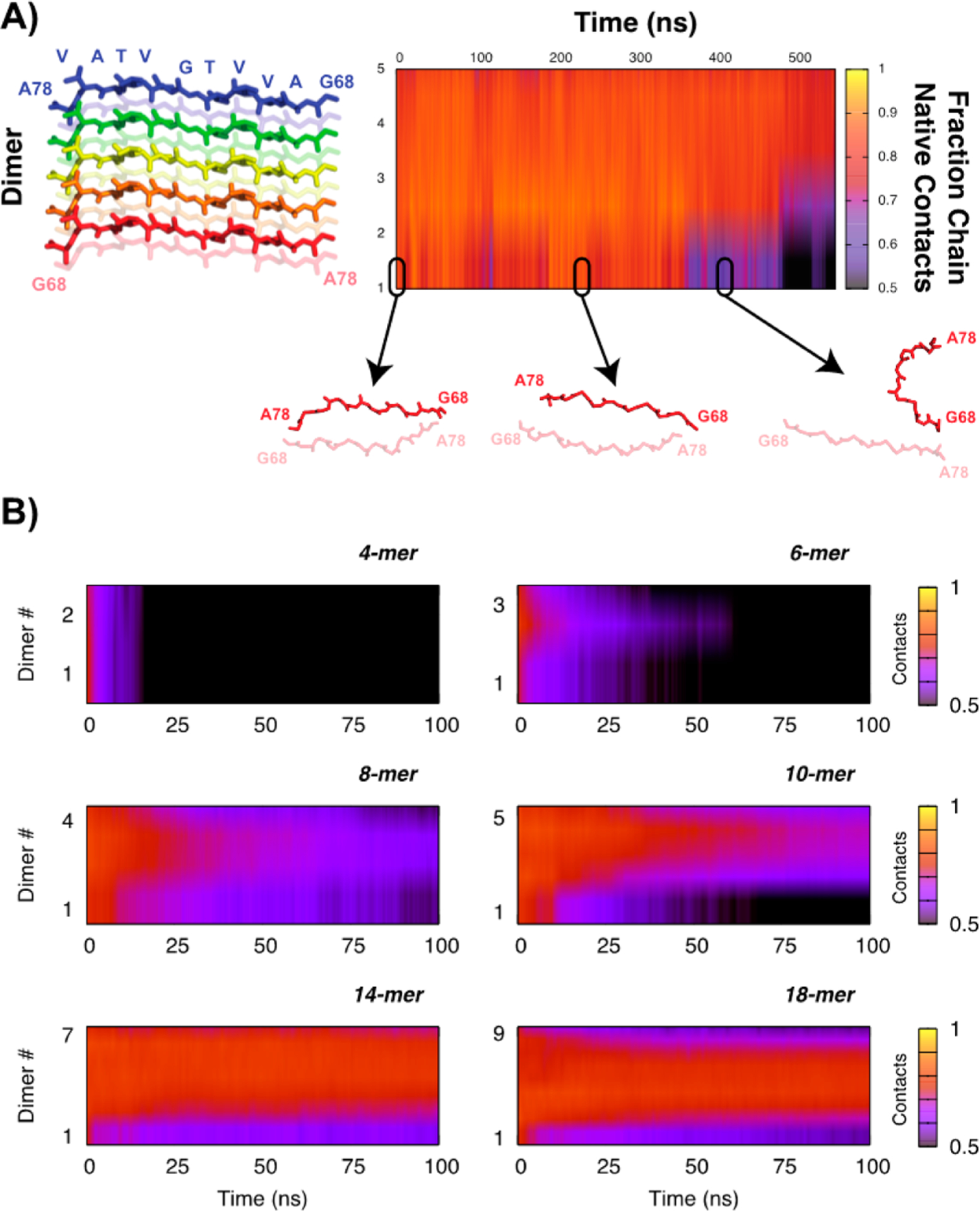
Figure 1.

(A) Data from a single 10-mer replica (5-dimers) showing the orientation of the dimers along the y-axis in the plot and representative structures for a single dimer depicting its dissociation and corresponding fractional native contacts. (B) Kinetic stability of the fibrils as a function of fibril size, averaged over 20 replicas. Similarity to the crystal structure is quantified using the average fractional native contact per dimer (1 indicates all native contacts are present, 0 means none of the contacts from the crystal structure are present).
