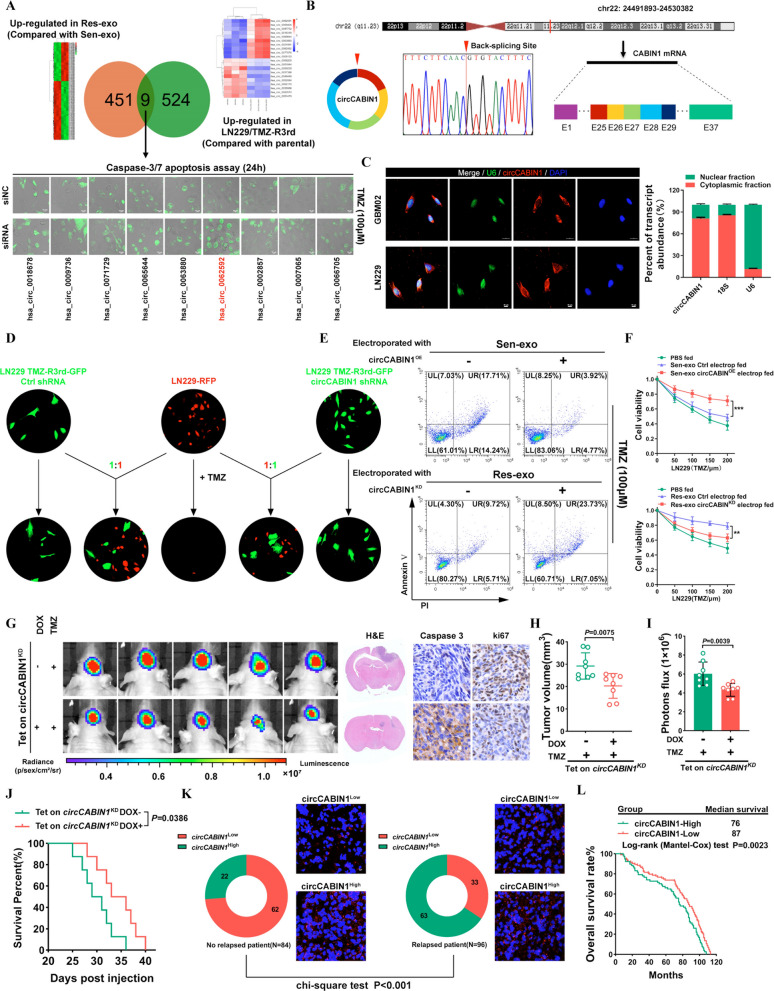
Fig. 2.
Intercellular transfer of circCABIN1 by exosomes disseminates temozolomide resistance. A Upper, Screening differentially expressed circRNA in TMZ-R cells and Res-exo by circRNA deep-sequencing. Lower, LN229 cells were transfected with siNC or siRNA and treated with TMZ (100 μM), and apoptosis was detected by caspase3/7 assay. Scale bar, 10 μm. B Explanation of the illustrated genomic loci of CABIN1, and the verification strategy for the circular exon 25–29 (circCABIN1). Sanger sequencing following PCR was used to show the “head-to-tail” splicing of circCABIN1 C Left, localization of circCABIN1 (red) in cells using fluorescence in situ hybridization (FISH). U6 probe coupled with Alexa Fluor™ 488 (green) and nuclei were counterstained with DAPI (blue). Scale bar, 10 μm. Right, nuclear and cytoplasmic fractions were isolated. circCABIN1 was mainly localized in cytoplasm. D TMZ-R3rd-GFP cells transfected with Ctrl shRNA or circCABIN1 shRNA co-cultured with parental cells (mCherry) at a ratio of 1:1 with TMZ treatment (100uM) for 4 days. Scale bar, 10 μm. E LN229 cells administrated with Sen-exo/Res-exo electroporated with circCABIN1OE/KD and TMZ were subjected to FACS to detect apoptosis. F The effect of Sen-exo/Res-exo electroporated with circCABIN1OE/KD on LN229 cells by CCK-8 assay. G-J Nude mice were orthotopically xenografted with Tet on circCABIN1 KD GBM cells (2 × 10⁶ cells) and treated with DOX-diet and intraperitoneally with TMZ (40 mg/kg) daily. Left, IVIS detects bioluminescence signals. Right, representative images of H&E and cleaved Caspase-3 and ki67 IHC in GBM sections of indicated groups. Quantification of tumor size (H), and quantification of bioluminescent imaging signal intensities (I), and survival rate (J) of mice in indicated groups. K Expression of circCABIN1 in relapsed or non-relapsed patients and representative immunofluorescence images in GBM section of indicated groups. L Kaplan–Meier analysis of OS in the high and low circCABIN1 groups according to the median circCABIN1 level in pre-therapy plasma (p = 0.0023). Results are presented as mean ± SD. **p < 0.01, and ***p < 0.001

