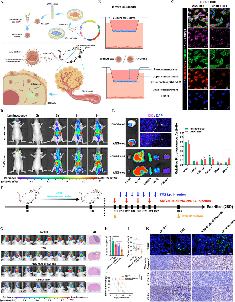
Fig. 7.
Targeted delivery of chemically modifed multi-siRNAs by engineered ANG-exo sensitized GBM cells to temozolomide. A Schematic image. Process of ANG-exo construction, isolation, multi-siRNA loading, animal tail vein injection. B Schematic image of the BBB model in-vitro. C Immunofluorescence images detected unmod-exo/ANG-exo uptake into LN229 cells after passing through a bEnd.3 monolayer. Scale bar, 10 μm. D In vivo florescence imaging of Orthotopic GBM xenograft mice at 0 h, 3 h, 6 h and 9 h time point after intravenous administration of unmod-exo / ANG-exo. E Left, ex vivo fluorescence images of Brain, Liver, Spleen, Lung, Heart, Kidney and frozen section of brain from mice sacrificed at 9 h post-injection. Right, fluorescence quantitative analysis of ex vivo organs of tumor-bearing mice after intravenous injection. F Schematic image. Time line of nude mice receiving combination therapy. G-J Verify the effect of combined therapy in vivo. IVIS detects bioluminescence signals. Quantification of bioluminescent imaging signal intensities (H), quantification of tumor size (I), and survival rate (J) of mice in indicated groups. K Representative images of TUNEL assay and cleaved Caspase-3, ALDH1A3 and OLFML3 IHC in GBM sections of indicated groups. Scale bar, 20 μm. Results are presented as mean ± SD. **p < 0.01 and ***p < 0.001

