Fig. 1.
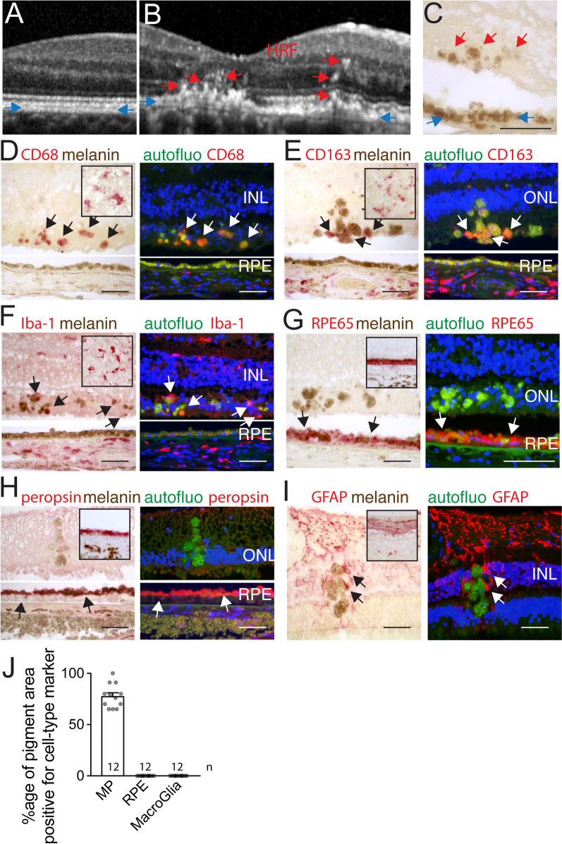
Intraretinal pigment in AMD is primarily located in melano-macrophages. RPE hyperreflective band (blue arrows) and hyperreflective foci (HRF, red arrows) visualized by SD-OCT of the retina of a healthy subject (A) and a patient with intermediate AMD (B). The aspect of the RPE (blue arrows) and retinal pigmented foci (red arrows) in an unstained paraffin section adjacent to the atrophic lesion of an AMD donor (C). CD68 (D), CD163 (E), IBA1 (F), RPE65 (G), peropsin (H), GFAP (I) staining in bright-field and fluorescence microscopy, of tonsils (insets C-F), healthy control retina (insets G-I) and retinal pigmented foci and adjacent RPE (C–I). The signal was revealed using Fast red chromogenic substrate visible in red in bright-field and in the red channel in fluorescence microscopy (arrows), autofluorescence was captured in the green channel and Hoechst nuclear stain in the blue channel. Immunohistochemistry experiments omitting the primary antibody served as negative controls (not shown). Calculation of the percentage of surface covered by immuno-stained retinal pigmented foci of total retinal pigmented foci for each immunostaining in each of the 12 donor eyes (J). HRF hyperreflective foci, RPE retinal pigment epithelium, INL inner nuclear lacer, ONL outer nuclear layer, scale bar = 50 µm; All values are reported as mean ± SEM

