Fig. 5.
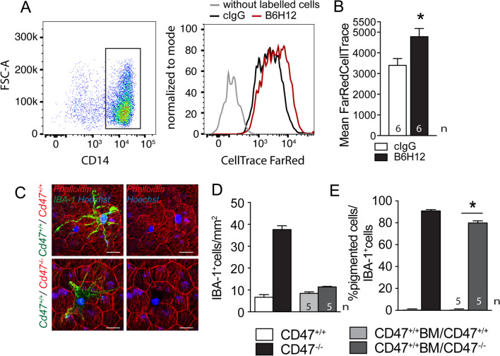
CD47-deficient RPE cells lose melanosomes/melanolipofuscin to melanophages. Gating and FarRed CellTrace intensity measurements by cytometry of human CD14 + Mo after 2 h of incubation with a monolayer of FarRed CellTrace pre-stained ARPE19 cells (a human RPE cell line) with 10 µg/ml of a control antibody (black line) or CD47 blocking antibody B6H12 (red line A) and quantification of the fluorescence intensity (B; n = 6 wells per group from three independent experiments; Mann–Whitney p = 0.0221). Three independent experiments gave similar results. Representative micrographs of phalloidin (red fluorescence staining), IBA1 (green fluorescence staining) double-labeled RPE/choroidal flatmounts of 12-month-old Cd47+/+ (upper panel) or Cd47−/−- recipient mice (lower panel) that had received a Cd47+/+- bone marrow transplant after lethal irradiation at 6 months of age (C). Quantification of the number of IBA1-stained subretinal MPs (D) and quantification of the percentage of pigment-laden melanophages (that block the phalloidin staining of the underlying RPE) of total subretinal MPs (E) of 12-month-old WT and Cd47−/−-mice compared with Cd47+/+ bone marrow transplanted WT and Cd47−/−-mice (n = 5/group; Mann–Whitney p = 0.00,159). Scale bar = 20 µm

