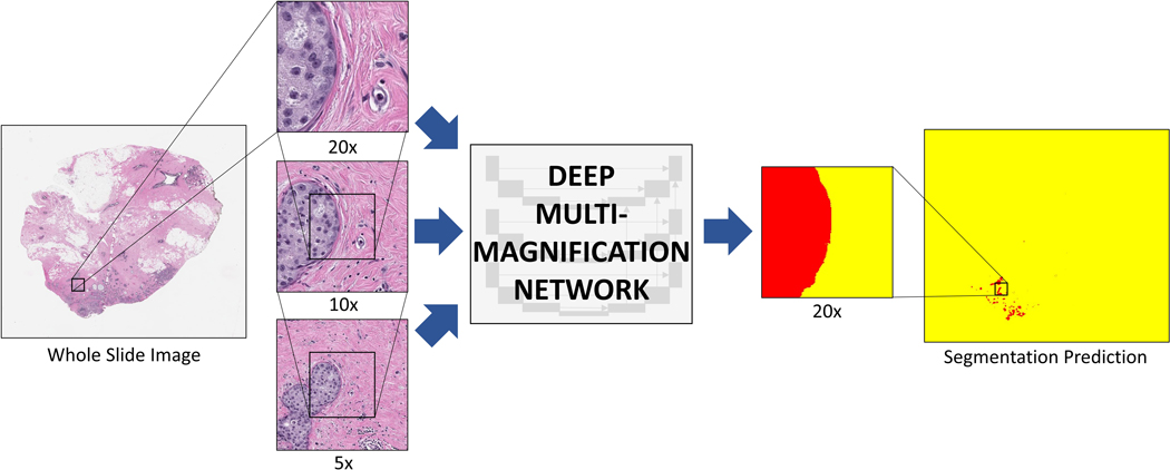
Figure 1. Deep multi-magnification network (DMMN).
Whole slide image (WSI) from a breast margin specimen with DCIS. The DMMN looks at a set of patches from multiple magnifications from the WSI allowing a wider field-of-view. The segmentation prediction image shows carcinoma highlighted in red, while the remaining tissue is highlighted yellow.

