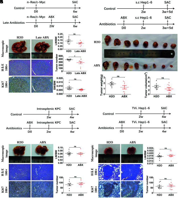
Fig. 2.
Gut commensal bacteria depletion does not accelerate liver tumor progression in mice. (A) Experimental procedure (n = 8 per group). (B) Representative macroscopic views of livers, H&E staining, and IHC of Ki67 in mouse HCCs. (C) The LW/BW ratio, tumor number, and the SW/BW ratio of each group. (D) Experimental procedure (n = 8 per group). Mice were treated with antibiotics or H2O for 2 wk before receiving subcutaneous injection of Hep1-6 tumor cell and, 12 d later, subcutaneous tumors were determined. (E) Macroscopic views of tumors. (F) Tumor weight and tumor volume of each group. (G) Experimental procedure (n = 9 per group). Mice were treated with antibiotics or H2O for 2 wk before receiving intrasplenic injection of KPC tumor cells and, 2 wk later, liver metastases were determined. (H) Representative macroscopic views of livers, H&E staining, and IHC of Ki67 in mouse liver metastases. (I) The LW/BW ratio and tumor number of each group were determined. (J) Experimental procedure (n = 10 per group). Mice were treated with antibiotics or H2O for 2 wk before receiving tail vein injection of Hep1-6 tumor cells and, 2 wk later, lung metastases were determined. (K) Representative macroscopic views of lungs, H&E staining, and IHC of Ki67 in mouse lung metastases. (L) The lung-body weight ratio (LW/BW) and tumor number of each group. Data were presented as mean ± SEM, P values were calculated by Student’s t test. ns, not significant; *P <0.05.

