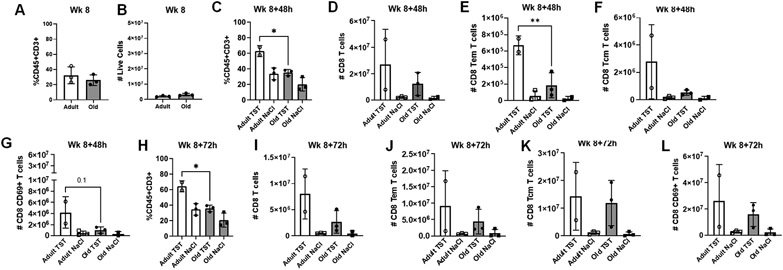
Figure 2. Adult macaques have increased T cells present in BALF following tuberculin instillation.
(A) Proportions of T cells (CD45+CD3+) in BALF of adult and aged macaques prior to tuberculin lung instillation (Week 8). (B) Number of live cells present in BALF of adult and aged macaques prior to tuberculin lung instillation (Week 8). (C) Proportions of T cells (CD45+CD3+) in BALF of adult and aged macaques 48 hours post-tuberculin challenge. (D) Number of CD8+ T cells, (E) CD8+ effector memory T cells, (F) CD8+ central memory T cells, and (G) CD69+CD8+T cells in BALF of adult and aged macaques 48 hours post-tuberculin challenge. (H) Proportions of T cells (CD45+CD3+) in BALF of adult and aged macaques 72 hours post-tuberculin challenge. (I) Number of CD8+ T cells, (J) CD8+ effector memory T cells, (K) CD8+ central memory T cells, and (L) CD69+CD8+ T cells in BALF of adult and aged macaques 72 hours post-tuberculin challenge. One-way ANOVA post-Tukey analyses comparing means between groups as indicated; *p<0.05, **p<0.01. TST = tuberculin instillation; NaCl = saline (control) instillation.

