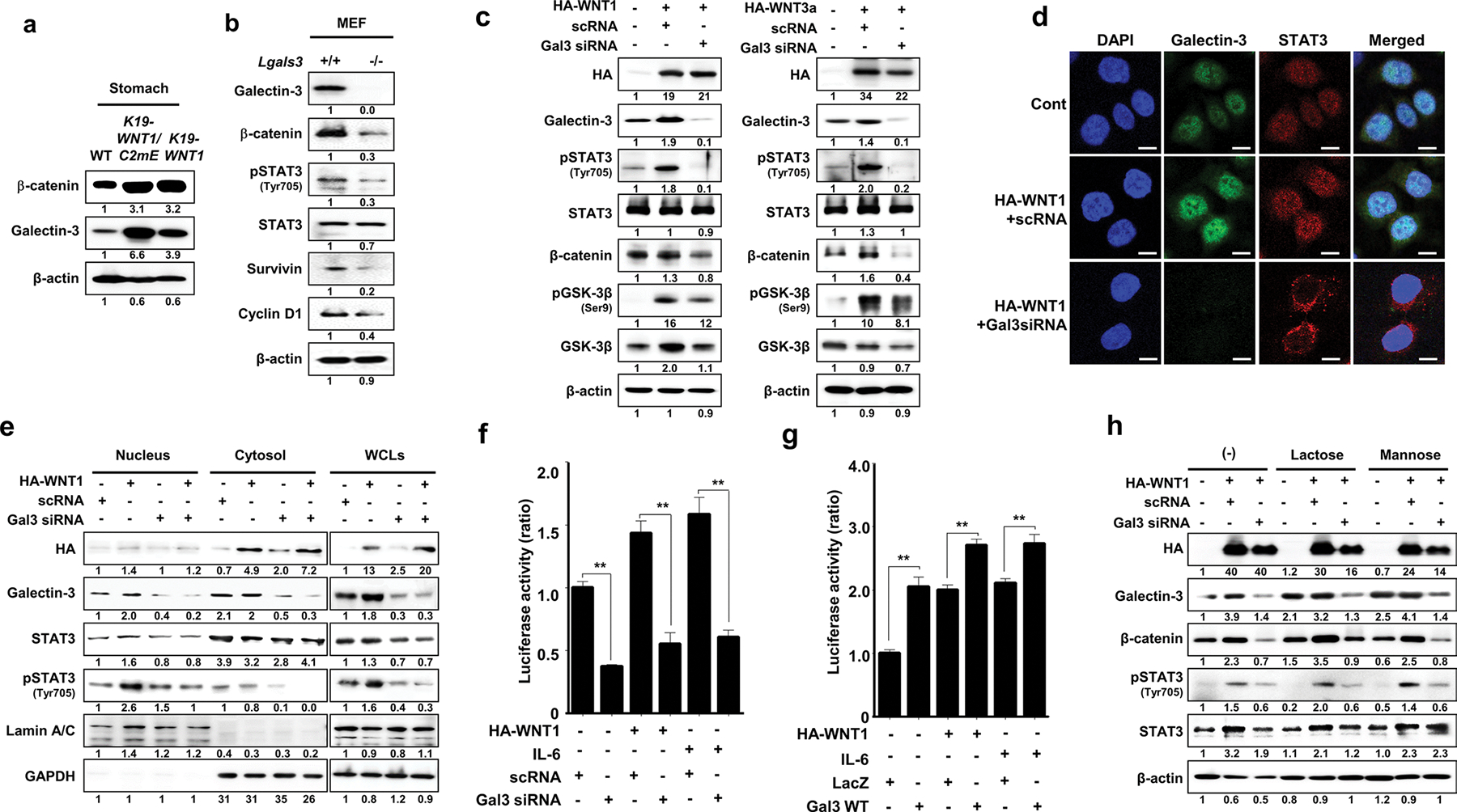
Fig. 2.

Overexpression of WNT1 induces phosphorylation and nuclear accumulation of STAT3 in a galectin-3-dependent manner. a Levels of β-catenin and galectin-3 in whole stomach tissues of WT mice and K19-WNT1/C2mE and K19-WNT1 transgenic mice. b Levels of the proteins indicated to the left in MEFs from WT and lgals3−/− mice. c Detection of the indicated proteins in AGS cells with or without transfection with HA-WNT1 (left) or HA-WNT3a (right) combined with galectin-3 silencing by siRNA. d Immunocytochemical analysis for the cellular localization of galectin-3 and STAT3 in WNT1-overexpressing and galectin-3-depleted AGS cells. Anti-galectin-3 FITC (green) and anti-STAT3 cy5 (red) antibodies were used, and cells were evaluated under a confocal microscope. DAPI was used to stain the nuclei (blue). Scale bars, 10 μm. e Detection of the indicated proteins in the nucleus, cytosol, or whole cell lysates of AGS cells with or without transfection with HA-WNT1 and galectin-3 siRNA. f, g STAT3 reporter luciferase activity in AGS cells transfected with HA-WNT1. At 24 h after plasmid transfection, the cells were treated with IL-6 with or without silencing of galectin-3 by siRNA (f) or overexpression of galectin-3 (g). The data are presented as the ratios of luciferase activities in untreated, non-HA-WNT1, and mock-transfected cells. Significant differences are indicated by asterisks (**p < 0.001). h Assay with the same experimental design as in the left of panel D, without or with a 4 h preincubation with 50 mM lactose or mannose before cell extraction
