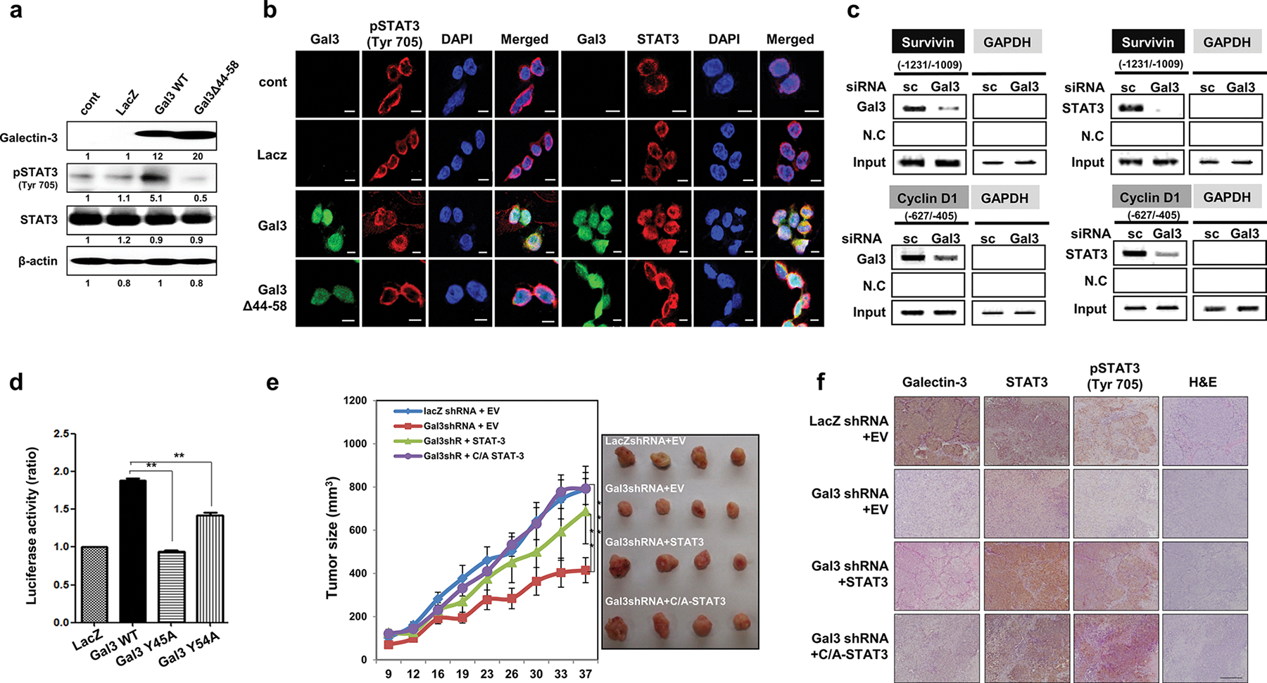
Fig. 5.

Galectin-3 increases the nuclear localization of STAT3 and regulates the DNA binding and transcriptional activation of STAT3. a,b SNU638 cells, which lack endogenous galectin-3, were infected with lentivirus vectors that overexpress LacZ, galectin-3 WT, and a STAT3 binding motif-deleted mutant of galectin-3 (Gal3Δ44–58). a The expression levels of galectin-3, STAT3, and pSTAT3 (Tyr705) as detected by western blotting. b Immunocytochemical analysis of the subcellular localization of FITC-galectin-3, Cy-5-STAT3, and Cy5-pSTAT3(Tyr705) in cells overexpressing LacZ, WT galectin-3, and Gal3Δ44–58. DAPI was used to visualize the nuclei (blue). Scale bars, 10 μm. Cells were fixed, and immunocytochemical analysis was performed using a confocal microscope. DAPI was used to visualize the nuclei (blue). Scale bars, 10 μm. c ChIP assays using antibodies against galectin-3 and STAT3 in AGS cells transfected with scRNA or gal3 siRNA. PCR fragments of the survivin and cyclin D1 promoters were detected. d SNU638 cells overexpressing lacZ, wild-type galectin-3 (Gal3 WT), and SH2 domain-binding motif mutant galectin-3 (Y45A or Y54A). This luciferase experiment was repeated three times, with similar results, and the data are shown as mean ± SD (n = 3). e AGS cells (106 cells) expressing LacZ, gal-3shRNA, gal-3shRNA, and wild-type STAT3, or gal-3shRNA and constitutively activated STAT3 (C/A STAT3) were subcutaneously inoculated into both flanks of a mouse to generate xenograft tumors (n = 5 per group). Tumor volume was measured for 37 days with calipers (as shown on the right). The error bars indicate the 95% confidence intervals (CI); *p = 0.0012, **p = 0.0023, and ***p = 0.0034, two-sided t test for values on the final day. f Galectin-3, pSTAT3(Tyr705), and STAT3 expression in mouse tumor tissues, as subcutaneously injected into the xenograft mouse model, was detected by IHC staining (brown), along with H&E staining. Magnification: 200×
