
Figure 6. Representative structures of the RBD-CRD interactions with KD in the structural ensemble of B-Raf autoinhibition in the absence of 14-3-3 dimer.
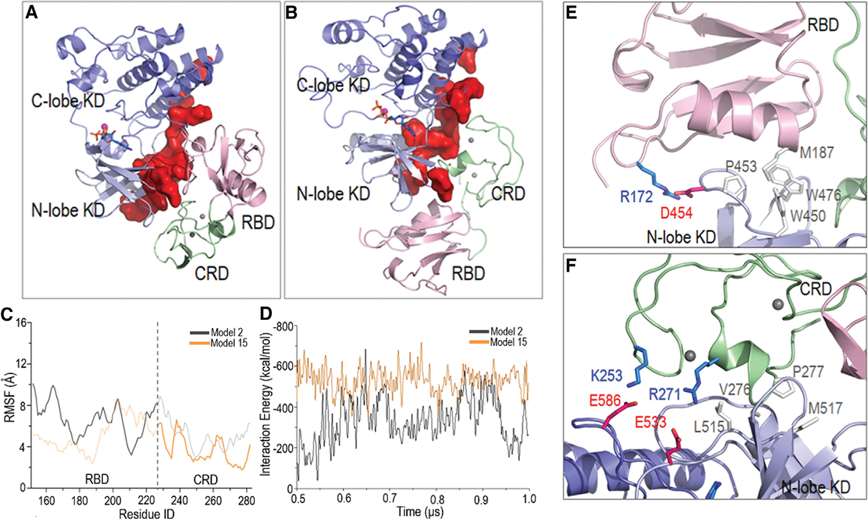
In (A) model 2 and (B) model 15, the KD dimer interfaces are largely blocked. The (C) root-mean-square fluctuations (RMSF) and (D) interaction energies indicate that model 15 is more stable. In RMSF curves, the RBD domain in model 2 and the CRD in model 15 that interact with the KD dimer interface are highlighted. In model 15, (E) the RBD and (F) CRD form salt bridges and hydrophobic interactions with the KD.