FIGURE 1.

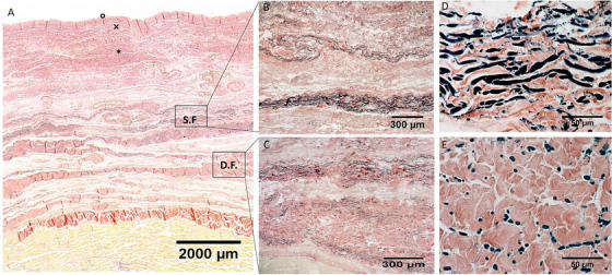
Histological images (Weigert Van Gieson staining) of anterior region (A), S.F. = Superficial Fascia; D.F. = Deep Fascia; × = papillary dermis; * = reticular dermis; ° = epidermis. (B) and (D): Superficial fascia. (C) and (E): Deep fascia

Histological images (Weigert Van Gieson staining) of anterior region (A), S.F. = Superficial Fascia; D.F. = Deep Fascia; × = papillary dermis; * = reticular dermis; ° = epidermis. (B) and (D): Superficial fascia. (C) and (E): Deep fascia