
FIGURE 3.
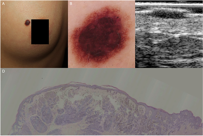
Compound nevus. (A) An elevated dark‐brown papule on the left breast. (B) Dermoscopic examination revealed central cobblestone and peripheral reticular pattern. (C) HF‐US at 20 MHz showed a typical straw‐hat sign. (D) Histopathology confirmed the dermal nevus cell aggregation at the lesion center (the cap under HF‐US), and the continuous band‐like nevus cell nests in the dermal‐epidermal junction at the lesion periphery (the brim under HF‐US), (hematoxylin‐eosin, original magnification ×25). HF‐US, high‐frequency ultrasound