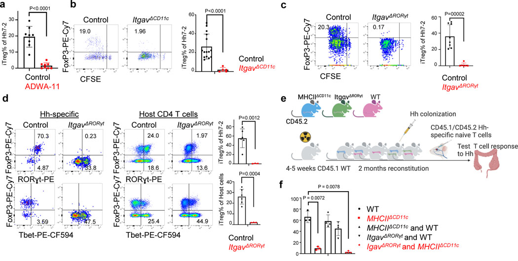
Figure 4. Role of integrin αvβ8 in RORγt+ antigen presenting cell-dependent iTreg cell differentiation.
a, Frequency of iTreg cells among proliferating donor-derived Hh-specific cells in the MLN at 3 days after transfer of naïve CFSE-labeled Hh7–2 T cells into mice treated with 200μg of ADWA11 blocking antibody (n=8) or left untreated (n=8), on the day of adoptive transfer. Data summarize three independent experiments. b, Hh7–2 T cell proliferation and differentiation in the MLN of ItgavΔCD11c (n=5) and littermate controls (n=15) at 3 days after adoptive transfer. Data summarize three independent experiments. c, Proliferation and differentiation of Hh-specific iTreg cells in the MLN of ItgavΔRORγt (n=6) and littermate control mice (n=8). CFSE-labeled Hh7–2 T cells were analyzed at 3 days following their adoptive transfer into Hh-colonized mice. Representative flow cytometry profiles (left) and aggregate data (right). Data summarize three independent experiments. d, Transcription factor expression in Hh7–2 T cells (left panels) and in endogenous CD4+ T cells (right panels) from colon lamina propria (LP) at 10 days after adoptive transfer into ItgavΔRORγt mice (n=3) and control littermates (n=6). Data summarize two independent experiments. Representative dot plots and aggregate data are shown (right panels). e, Scheme for mixed bone marrow chimeric mouse experiment, with control, ItgavΔRORγt or MHCIIΔCD11c cells administered to irradiated host mice (created with BioRender.com). f, Bar graphs showing iTreg frequency among Hh7–2 T cells in the colon LP at 10 days after their transfer into the bone marrow chimeric mice, reconstituted with different combinations of donor cells as indicated. Control mice (n=3), MHCIIΔCD11c (n=3), MHCIIΔCD11c and WT (n=4), ItgavΔRORγt and WT (n=4) and MHCIIΔCD11c and ItgavΔRORγt (n=4). All statistics were calculated by unpaired two-sided Welch’s t-test. Error bars denote mean ± s.d. p-values are indicated in the figure.

