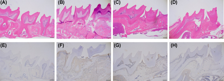
Figure 3. H&E staining and immunohistochemical localization of IL-6.
H&E staining of periodontal tissues in the control group (A), ligature-induced periodontitis (B), recovery for 14 days (C), recovery for 28 days (D), IL-6 expression in periodontal tissues of control mice (E), and elevated expression of IL-6 in periodontal tissues of mice with ligature-induced periodontitis (F), recovery for 14 days (G), recovery for 28 days (H). Magnification: 40×; scale bar: 200 μm.

