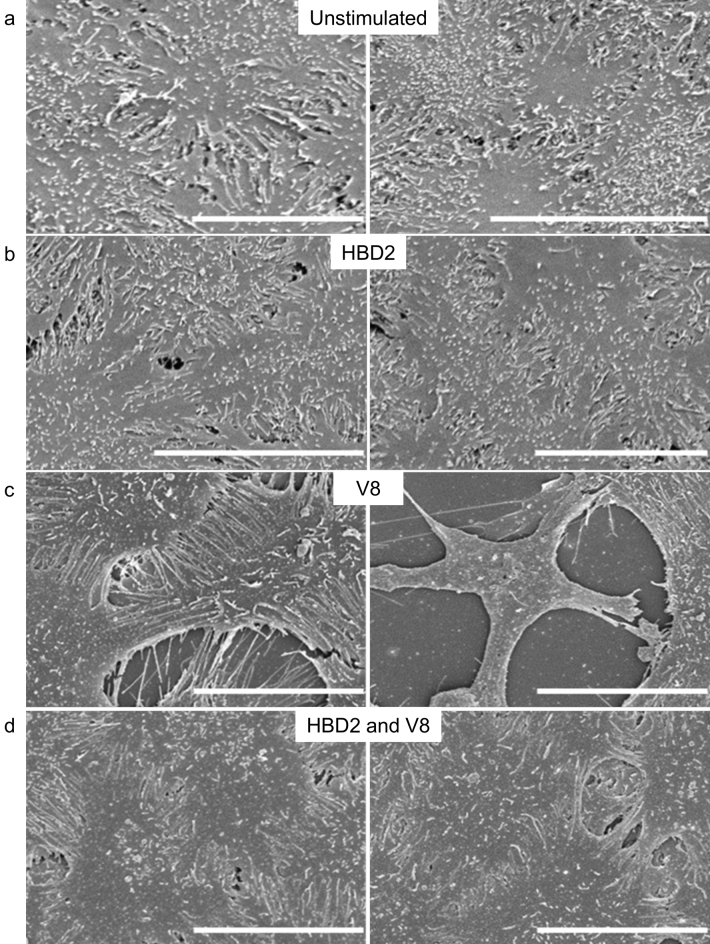
Figure 4.
Scanning electron microscopic imaging of HaCaT cells exposed to HBD2 and/or V8. HaCaT cells were seeded into 35 mm corning coated TC dishes. Once confluency was reached, cells were treated with vehicle control (a,c) or 0.5 μg/ml synthetic HBD2 (b,d). After 24 h, cells were washed and then exposed to vehicle control (a,b) or 5 μg/ml recombinant V8 (c,d) for an additional 24 h, before cells were fixed with PFA and glutaraldehyde buffer overnight at 4 °C. Cells were then washed; glutaraldehyde buffer was added for a further overnight incubation at 4 °C and prepared for imaging. Samples imaged with the Hitachi S-4700 scanning electron microscope. Scale bars represent 20 μm. Images representative of 5 images taken per condition.

