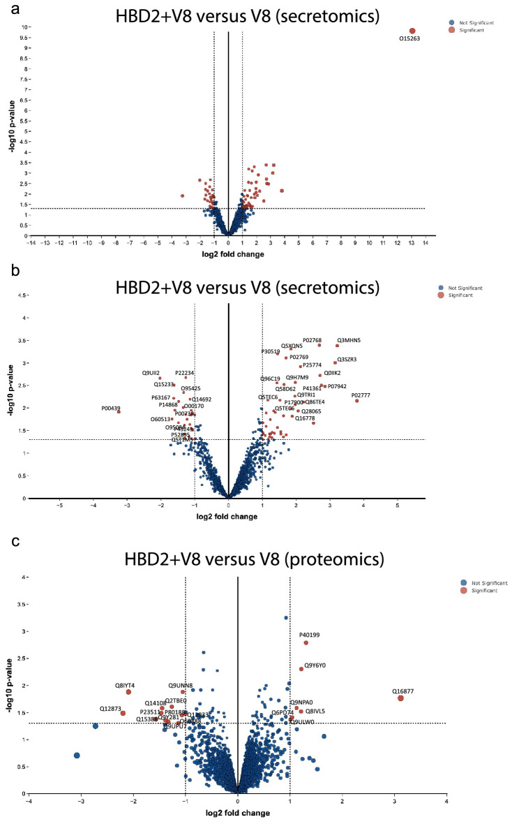
Figure 6.
Secretomic and proteomic characterisation of HBD2-mediated protection from V8 damage in HaCaT cells. HaCaT cell monolayers were treated with 0.5 μg/ml HBD2 in serum free media for 24 h, before addition of 5 μg/ml V8, or vehicle control, for a further 24 h. Supernatants (a,b) and cells (c) were then collected, centrifuged at 3000 rpm for 5 min, and stored at -80 °C prior to analyses. n = 5 per condition. Log fold changes and p values generated using LIMMA pathway43. (a,b) Volcano plots generated from secretomics analysis from the supernatants of HaCaT cells protected with HBD2 before exposure to V8, compared to unprotected V8-damaged cells. Red points indicate significant points, blue indicate nonsignificant points. (a) Data represented as a whole. (b) Data represented without O15263 data point, zoomed in to central portion of graph. (c) Volcano plot generated proteomics analysis of HaCaT cells protected with HBD2 before exposure to V8, compared to unprotected V8-damage cells. Red points indicate significant points, blue indicate nonsignificant points.

