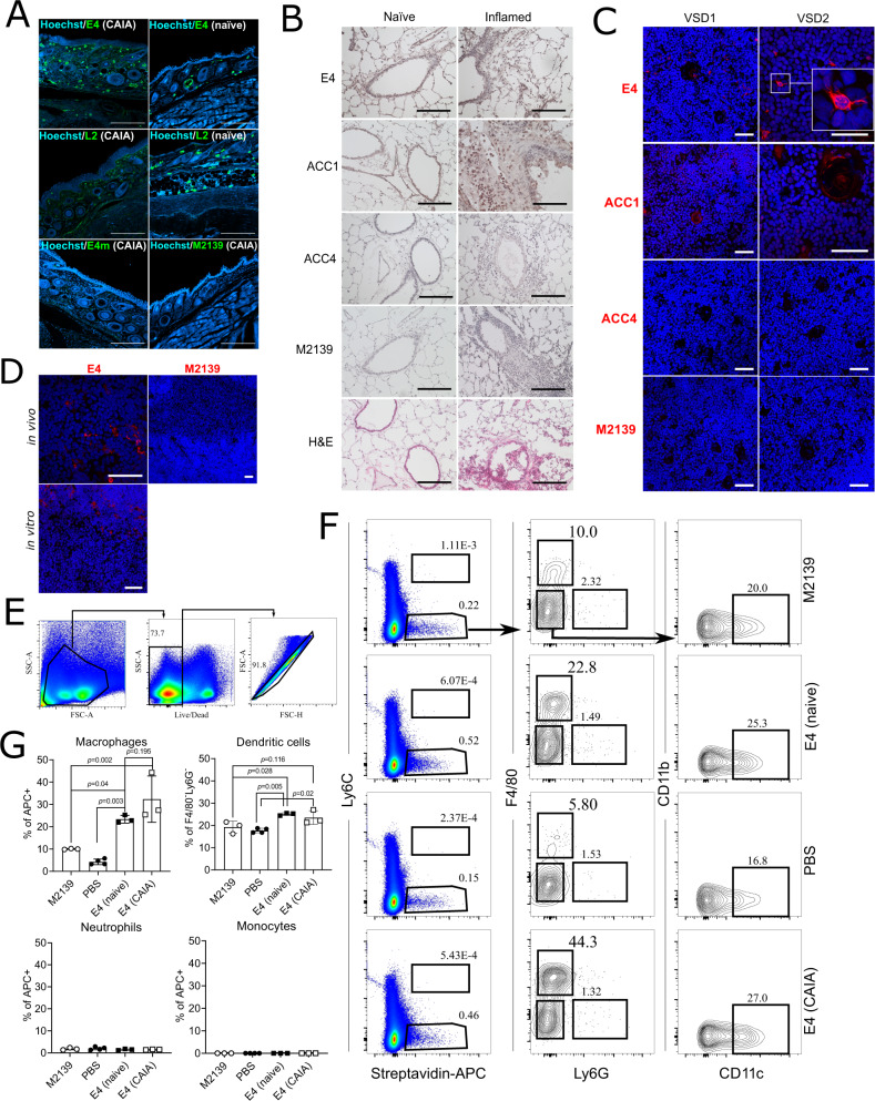
Fig. 4. Reactivity of E4 to non-joint tissues/cells.
A E4 binding to skin tissues from naïve or CAIA arthritic joints. Immunofluorescence (IF) staining was performed using the indicated mIgG2b antibodies on skin tissue from naïve or arthritic joints from DBA/1 mice with CAIA (day 15), the antibodies were detected using a goat anti-mouse IgG secondary antibody conjugated with CF488A and DNA was stained with Hoechst. Images were captured by confocal microscopy at 10× magnification. Scale bars represent 200 µm. B E4 binding to naïve and inflamed lung tissue. IHC/H&E staining was performed using the indicated antibodies on either healthy or mannan-induced inflamed lung tissue taken from B10.Q mice. Images were captured by light microscopy at ×20 magnification and scale bars represent 100 µm. C E4 binding to human and (D) murine thymus tissue from B10.Q mice. Immunofluorescence (IF) staining of the thymus from two individuals or mice was performed using the indicated biotin-labeled antibodies labeled. For in vivo binding, biotinylated antibodies were pre-injected. Antibodies and cell nuclei were visualized by Streptavidin Alexa Fluor 555 (red) and Hoechst33342 (blue), respectively. Images were captured by confocal microscopy at ×10, ×20 or ×40 magnification. Scale bars represent 50 µm. E–G The binding of E4 to splenocytes in vivo. Splenocytes from naïve or CAIA arthritic mice injected with the indicated biotinylated antibodies (n = 3 or 4) were analyzed by flow cytometry. E, F Represent the general gating strategy. Macrophages are in the F4/80+Ly6G− population gated on streptavidin-APC+ cells, dendritic cells in the CD11c+CD11b− population gated on F4/80−Ly6G− cells, monocytes in the Ly6C+streptavidin-APC+ population, and neutrophils in the F4/80-Ly6G+ population gated on streptavidin-APC+ cells. Data are assessed by Mann–Whitney test (two-tailed) and presented in (G) as mean ± SD.

