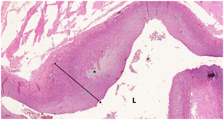
Figure 1.
Photomicrograph of the hepatic artery wall in the hilum of a liver obtained from a participant with alcoholic cirrhosis. Atherosclerosis and arterial intimal thickening is apparent, with a >10% reduction in the diameter of the arterial lumen (L). The line defines the distance between the vascular endothelium (★) and the internal elastic limit (arterial intimal layer) (●), and × indicates foam cells. The scale bar (lower left corner) represents 200 µm. Hematoxylin and eosin, 40× magnification.

