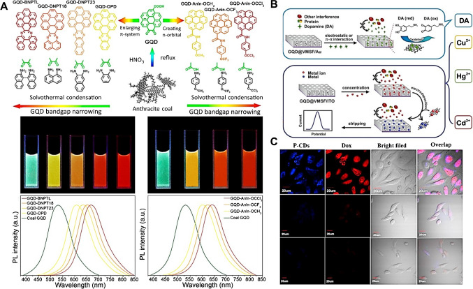
Fig. 2.
(A) (Top) Schematic representation of the tuneable photoluminescence redshift caused by the introduction of n-orbitals or by the enlargement of π-systems in GQDs, with their photographs under a UV lamp (middle) and the respective spectra (bottom). Reproduced from [38] with permission from the American Chemical Society. (B) Incorporation of GQDs into a mesoporous silica–nanochannel film through electrophoresis, resulting in anti-fouling properties for the electrochemical sensing of Hg2+, Cd2+, Cu2+, and dopamine (DA) in complex samples, such as food, soil, and serum. Reproduced (adapted) from [48], with permission from the American Chemical Society. (C) Bioimaging of HeLa cancer cells based on the fluorescence of CQDs after endocytosis due to preferential hyaluronic acid–CD44 interactions compared to normal cells, in blue and red channels, and the corresponding bright field (first row); HeLa cancer cells pre-treated with hyaluronic acid, showing weak PL (middle row), and non-cancer NIH-3T3 cells indicating weak PL (bottom row). Reproduced from [49], with permission from Elsevier

