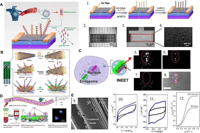
Fig. 3.
(A) (Left) schematic of the CNT-FET based biosensing platform for the detection of breast cancer exosomal miRNA21, in an example of affinity sensing relying on CNTs and a designed probe DNA. The probe is built by (1) the incorporation of gold nanoparticles and thiolated DNA; (2–3) SEM images of the biosensor chip. (4) Part of the device channel with AuNPs uniformly distributed on the Y2O3/CNT film. Reprinted (adapted) from [67] with permission from the American Chemical Society. (B) Representation of the electrochemical sensing pad designed by the incorporation of gold nanocages (AuNCs) and probe DNA onto MWCNTs; methylene blue adsorbed after the sensor’s exposition to the sample with the target DNA. Reproduced from [70], with permission from Nature. (C) Application of CNT-based FRET nanoprobes to the bioimaging of sub-cellular structures as (5, 6) the nuclear region and (7) cytosolic region, generating (8) a combined artificially colored image through multiplexed spectral analysis. Reprinted (adapted) from [71], with permission from the American Chemical Society. (D) Operational schematic for the CNT-based antigen-independent capture of circulating tumor cells in human blood serum, exploiting the preferential interaction of CTCs to CNTs and enabling the observation of multiple phenotypes of breast cancer cells. Reproduced from [72] with permission from the Royal Society of Chemistry. (E) (9) FEG–SEM image of the flexible carbon fibers with graphene oxide exfoliated directly onto the filaments’ surface, (10) cyclic voltammograms of FCF and FCF-GO and (11) FCF-GOx and FCF-GO-GOx in sodium phosphate buffer, (12) oxidative electron-transfer rate constant as a function of the overpotential for FCF-GOx and FCF-GO-GOx. Reproduced from [73], with permission from the Royal Society of Chemistry

