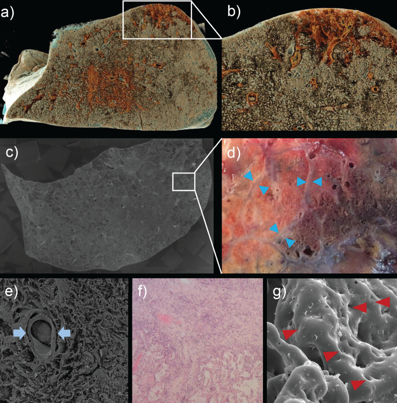
FIGURE 1.
a) Cinematic rendering of a hierarchical phase-contrast tomography (HiP-CT) study from a 78-year-old male patient who succumbed to COVID-19 highlights the spatial heterogeneity of the affected lung parenchyma. b) Close-up of patchy subpleural consolidations reveals the heterogenous distributions of functional alveolar dead space in COVID-19 patients. c) HiP-CT of a COVID-19 lung imaged at 25 µm per voxel depicts the mosaic distribution of secondary pulmonary lobules with pulmonary microvascular involvement and occlusions. d) Gross appearance of an upper lobectomy of a 62-year-old patient with post COVID conditions (6 months after acute COVID-19 pneumonitis) demonstrating the spatial heterogeneity of consolidated secondary pulmonary lobules. Interlobular septa with a thickness of approximately 0.1 mm (blue arrowheads). e) Scanning electron micrograph revealed the complete occlusion of a centrilobular artery (blue arrows) and f) thickening of interlobular septae (haematoxylin and eosin-stained section). g) Secondary lobular micro-ischaemia in long COVID results in an even, prolonged blood vessel neo-formation by intussusceptive angiogenesis (red arrowheads).

