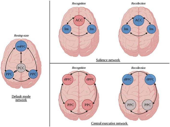
FIGURE 2.
Representation of the neural correlates of dissociative amnesia (DA). Neuroimaging studies of DA show that patients had several differences in their brain functioning compared to healthy subjects. In a resting state condition, through the default mode network, they had a decreased activation of the ventromedial prefrontal cortex and the bilateral posterior parietal cortices. Abnormal patterns were also identified during recognition and recollection tasks in both salience and central executive networks. ACC, anterior cingular cortex; dlPFC, dorsolateral prefrontal cortex; Ins, Insula; PCC, posterior cingular cortex; vmPFC, ventromedial prefrontal cortex. Blue circles represent hypoactivations. Red circles represent hyperactivations. Gray circles represent normal functioning.

