Figure2 .
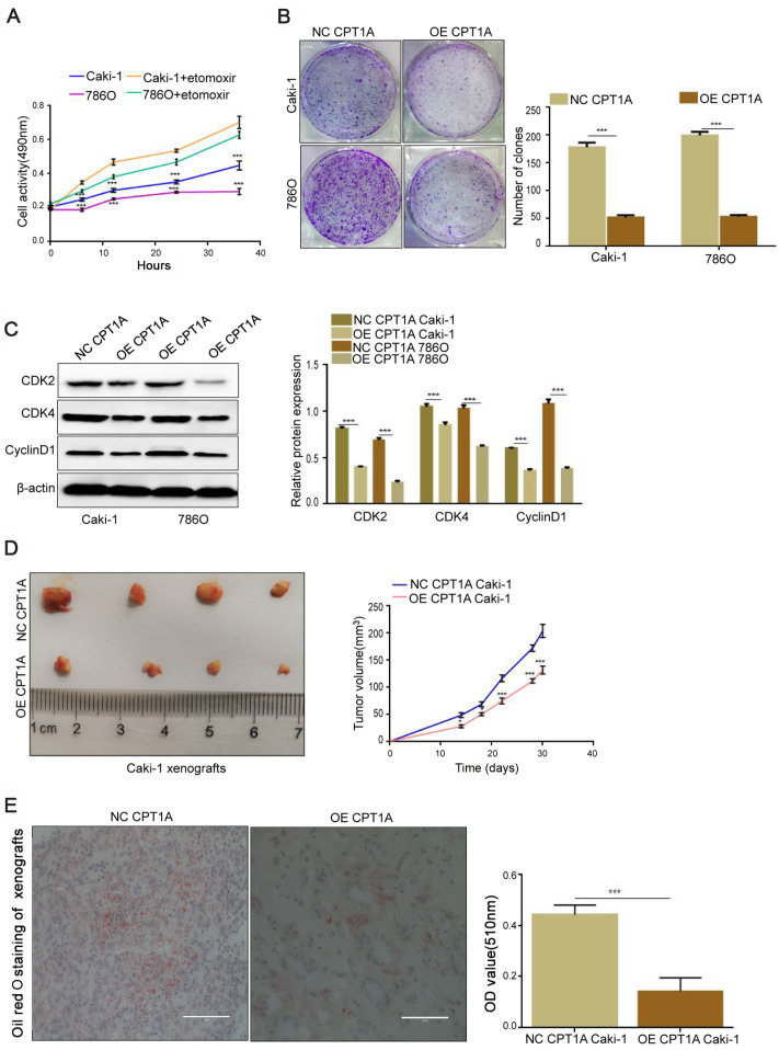
CPT1A overexpression prevents lipid accumulation and cell proliferation in vivo and in vitro (A) Cell proliferation of Caki-1 and 786O incubated with etomoxir (80 μM) at 0, 6, 12, 24, and 36 h detected by MTS assay. (B) Colony formation assay of Caki-1 and 786O stably transfected with CPT1A overexpression lentivirus indicated that overexpression of CPT1A could inhibit the proliferation in ccRCC. (C) The expressions of CDK2, CDK4 and CyclinD1 detected by western blot analysis. (D) Orthotopic xenograft transplanted with NC (n = 6) and OE-CPT1A/Caki-1 cells (n = 6). Tumor volume at day 14 was measured in each group. (E) Oil red O staining of frozen sections of tumor tissue from nude mice. Scale bar = 50 μm. Values are shown as the mean±SD. *P<0.05, ***P<0.001 (Student’s t-test).

