Figure2 .
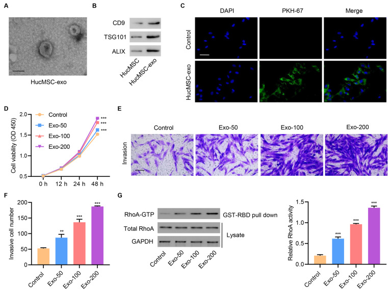
HucMSC exosomes increase cell proliferation, invasion, and RhoA activity in primary injured tenocytes(A) A typical electron microscopy image of exosomes from hucMSCs. Scale bar=100 nm. (B) Representative western blots of CD9, TSG101, and ALIX proteins. (C) Laser scanning confocal microscopy was used to visualize exosome internalization by primary injured tenocytes. Scale bar=50 μm. Injured tenocytes were treated with different concentrations of hucMSC exosomes (50, 100, and 200 μg/mL), and (D) cell proliferation (E,F) invasion, and (G) RhoA activity were determined. Scale bar=100 μm. Data are expressed as the mean±SD (n=3). **P<0.01, ***P<0.001 compared with the control.

